Cortical Biopsies Hint at Start of Alzheimer's 'Cellular Phase'
Quick Links
What goes on inside the brain of a person in the preclinical stages of Alzheimer’s disease? Science grapples with this question by studying postmortem tissue, despite concerns about agonal changes and postmortem interval degrading sample quality. Now, a unique collection of surgical biopsies offers a fresh perspective. In a manuscript uploaded June 5 to bioRxiv, researchers led by Evan Macosko and Beth Stevens at the Broad Institute of MIT and Harvard report the first single-nucleus RNA-Seq analysis of cortical samples that were were taken during surgery to implant a ventricular shunt to relieve symptoms of hydrocephalus.
- Surgical biopsies offer a rare peek at AD pathology in living brain.
- Inhibitory neurons are lost, excitatory neurons are transiently hyperactive.
- Inflammatory microglia proliferate.
- Neurons and oligodendrocytes make more Aβ.
Integrating this new biopsy data with prior postmortem transcriptomic data, the scientists compiled an enormous set of cell profiles spanning disease states and species. From this emerged a cellular signal of early AD, in living people, that has never been seen before.
It reveals loss of a particular set of layer 1 inhibitory neurons along with transient hyperactivation of a particular set of layer 2/3 excitatory neurons. Amyloidogenic APP processing ramps up in pyramidal neurons and oligodendrocytes, and a specific cluster of microglia expands. The dataset offers a glimpse into the cellular phase of AD, a years-long debacle that unfolds after amyloid forms and during which malfunctioning neurons, glia, and endothelial cells damage the brain (De Strooper and Karran, 2016).
“What sets this data apart is that we found some credible biological insight into early Alzheimer’s,” Macosko told Alzforum. “We have seen clear evidence of the hyperactivity hypothesis in AD brain.”
“This exciting study [...] stands out methodologically,” noted Martin Kampmann, University of California, San Francisco. Rick Livesey, University College London, cautioned that the donors' normal-pressure hydrocephalus may complicate interpretation of the results, but called the overall work "a welcome and insightful contribution to understanding the cell and molecular biology of dementia in humans in vivo."
Like many a good collaboration, this study was born at a conference, Macosko told Alzforum. Wanting to run large-scale transcriptomics, but concerned about the limitations of postmortem tissue, Macosko wished for a cohort of good ex vivo samples. Henrik Zetterberg, from the University of Gothenberg, Sweden, knew just whom to ask. He introduced Macosko and Stevens to Ville Leinonen, a neurosurgeon at the University of Eastern Finland, in Kuopio. For years, Leinonen has been collecting, storing, and characterizing samples of frontal cortex taken during hydrocephalus shunt placements (see Parts 1 and 2 of this series).
“We have worked with other surgical samples before, but Ville’s attention to detail is unparalleled, and he has a personal interest in obtaining the highest quality data,” said Macosko.
With tissues biopsies from 52 donors, frozen within five minutes of coming out of the brain, joint first authors Vahid Gazestani and Tushar Kamath and colleagues used single-nucleus RNA-Seq to profile the transcriptomes of nearly 900,000 cells, or 17,000 per donor. Nineteen samples had amyloid plaques, eight had plaques and evidence of phosphorylated tau, 25 had neither. By clustering like transcriptomes, the scientists identified the major cell types: excitatory and inhibitory neurons, microglia, astrocytes, endothelial cells/pericytes, oligodendrocytes, and oligodendrocyte precursors. Further clustering within each produced 82 subtypes.
The scientists reasoned that with this high-quality dataset, they'd have enough power to integrate and characterize previous data with greater specificity. From 27 published studies, almost 2.5 million cells met quality-control criteria and were annotated based on the 82 cell subtypes identified in the Kuopio cohort. The prior analyses were of human postmortem samples of people with AD, Parkinson’s, multiple sclerosis, and autism, plus datasets from mouse models of AD and related disorders.
What did this analysis reveal? First, the comparison of fresh to postmortem tissue confirmed that the morbidity just before death, and degradation after, had indeed changed gene-expression patterns in the prior datasets. For example, it rendered inhibitory and excitatory neurons less complex, and generated expression artefacts in microglia.
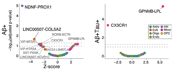
How about an AD signature? By correlating gene expression with pathology burden, Gazestani and colleagues recognized cellular changes specific to preclinical AD. First, they noticed that two specific types of neuron were depleted in cortex with amyloid but not tau pathology. They are interneurons expressing the genes NDNF and PROX1, and excitatory neurons expressing LINC00507 and COL5A2. These neurons lie in layer 1 and layers 2/3 of the cortex, respectively.

Early Losses. In frontal cortices from living people who have only amyloid pathology, two groups of neurons are depleted: those expressing NDNF-PROX1, and those expressing LINC00507-COL5A2 (left). In people who have both amyloid and tau pathology, there are more microglia expressing GPNMB and LPL genes, and fewer microglia expressing CX3CR1. [Courtesy of Gazestani et al., bioRXiv, 2023.]
In people with high amyloid pathology plus tau phosphorylation, the relative dearth of these neurons was no longer evident. Macosko suspects this is because by this later stage of pathology, other neurons start dying, too.
Are these cellular changes coordinated? A look at gene-expression patterns in the excitatory neurons suggests as much. The authors found two pools of differentially expressed genes, aka reactomes, in these cells. One emerged in cortices that had only amyloid pathology, and petered out in cortices that had both amyloid and tau pathology. The second surfaced once both pathologies were present.
Gene set enrichment analysis identified pathways behind these reactomes. When amyloid burden was present, the layer 2/3 LINC00507-COL5A2 excitatory neurons ramped up genes needed for glucose metabolism, the tricarboxylic acid cycle, and mitochondrial electron transport—as if they were trying to meet demand for more energy. When amyloid burden was high and phosphorylated tau had entered the picture, expression of these same genes waned, suggesting the neurons were struggling to survive. Cortical neurons in deeper layers mounted much weaker gene-expression changes, another indication that this particular neuronal response might be quite specific (image below).
Layer 2/3 LINC00507-COL5A2 excitatory neurons also transiently activated cell protective genes, e.g., those for cholesterol synthesis, scavenging reactive oxygen species, DNA repair. Tellingly, loss of the L1 NDNF-PROX1 inhibitory neurons correlated with these expression changes. In addition, the fewer inhibitory neurons were in the sample, the more the excitatory neurons ramped up genes typically induced after neural activity, such as FOS and JUN. This hints that the loss of this L1 inhibition increases activity in L2/3 excitatory neurons.

Spot the Reactome. Columns show types of excitatory neurons, each with three levels of increasing AD pathology from left to right. Upper layer LINC00507-COL5A2 and RORB_SCTR neurons respond (Y axis) to amyloid and tau pathology, but only transiently. Expression of genes needed to meet energy demands (rows, see glucose metabolism, TCA cycle, electron transport) turns up (red dots) when amyloid pathology (x axis) is present, but peters out (blue dots) when amyloid burden is high and tau is phosphorylated. Reactomes protecting against reactive oxygen species and DNA damage are also transiently activated. Lower layer neurons do not react this way. [Courtesy of Gazestani et al., bioRXiv, 2023.]
“Collectively, our results demonstrate NDNF-PROX1 inhibitory neuron loss is correlated with hyperactivity and preferential loss of layer 2/3 excitatory neurons in the prefrontal cortex with low Aꞵ plaque burden,” the authors concluded. Scientists have long known of paradoxical hyperactivation in early AD and animal models, and evidence has implicated loss of inhibitory input (Palop et al., 2007; Busche et al., 2008; Putcha et al., 2011; Verret et al., 2012).
Gazestani et al. call their finding the Early Cortical Amyloid Response. It has additional components. Take the 400,000 microglia in this study. Their transcriptomes fell into 13 subtypes, of which two—marked by LPL/CD83 and GPNMB/EYA2 expression—were becoming relatively more numerous with increasing pathology. The latter subtype also behaves this way in PD, and in mouse models of ADRD, ALS, and multiple sclerosis, suggesting these microglia represent a general response to pathology or neurodegeneration. Expansion of LPL/CD83 microglia appears specific to AD.
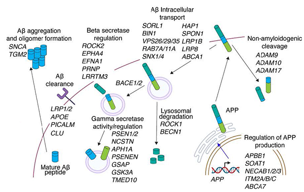
Another change jumped out. When there were plaques, genes responsible for Aβ production, including APP itself, were turned up. The authors were surprised to see this not only in the hyperactive excitatory neurons, but also in oligodendrocytes. For both cell types, the signature was strongest in samples with the lowest amyloid burden, suggesting a response to early pathology. Genes known to temper Aβ production, including SORL1 and PICALM, were down (image below).

Trouble Brewing. According to analysis of a set of 45 genes involved the production of Aβ, hyperactive neurons and oligodendrocytes in living people with AD pathology make more of the peptide. [Courtesy of Gazestani et al., bioRXiv, 2023.]
Macosko thinks perhaps the oligodendrocytes ramp up Aβ production because they are trying to assist the neurons. “APP production as a fraction of total expression is higher in oligodendrocytes than in neurons, so the glia are putting a lot of energy into making it,” noted Macosko. “I think the uptick is probably non-autonomous. It may be a response to the hyperactive neurons, but we don’t know that yet."
Are these early changes a response to amyloid burden? That’s the assumption, but Macosko said it is still a mystery. Unclear also: whether the loss of inhibitory neurons precedes, or causes, the hyperactivation and loss of excitatory neurons. “Right now, all we have is a correlation,” he said.
Neither does he know why these particular neurons are affected. The scientists searched for shared characteristics that might explain their vulnerability, such as ion channels or patterns of activity or expression. Nothing stood out. Perhaps these neurons' position in the outer layers, closer to the brain border, may expose them more to cytokines and other molecules that might pose a threat. Small samples of dura are also being collected during shunt surgery, and starting to be studied.
Or perhaps the neuron's circuitry makes them vulnerable to stress because they receive inputs from relatively far away. Further studies might tell. “We now have a system where we can probe these cells electrophysiologically in acute slices to ascertain their excitability and transcriptional response. That opens the door to exciting opportunities,” he said (see Part 3 of this series).
Meanwhile, the biopsy donors live on, as participants in a longitudinal NPH research program.—Tom Fagan and Gabrielle Strobel
References
News Citations
- Fresh Brain Every Friday: Biopsies Transform Alzheimer's Science
- A Day's Work: Cortex Biopsy Comes Out. Shunt Goes In. Patient Goes Home.
- Brain Tissue From Living People with Amyloid Plaques Can Fire in a Dish
Paper Citations
- De Strooper B, Karran E. The Cellular Phase of Alzheimer's Disease. Cell. 2016 Feb 11;164(4):603-15. PubMed.
- Palop JJ, Chin J, Roberson ED, Wang J, Thwin MT, Bien-Ly N, Yoo J, Ho KO, Yu GQ, Kreitzer A, Finkbeiner S, Noebels JL, Mucke L. Aberrant excitatory neuronal activity and compensatory remodeling of inhibitory hippocampal circuits in mouse models of Alzheimer's disease. Neuron. 2007 Sep 6;55(5):697-711. PubMed.
- Busche MA, Eichhoff G, Adelsberger H, Abramowski D, Wiederhold KH, Haass C, Staufenbiel M, Konnerth A, Garaschuk O. Clusters of hyperactive neurons near amyloid plaques in a mouse model of Alzheimer's disease. Science. 2008 Sep 19;321(5896):1686-9. PubMed.
- Putcha D, Brickhouse M, O'keefe K, Sullivan C, Rentz D, Marshall G, Dickerson B, Sperling R. Hippocampal hyperactivation associated with cortical thinning in Alzheimer's disease signature regions in non-demented elderly adults. J Neurosci. 2011 Nov 30;31(48):17680-8. PubMed.
- Verret L, Mann EO, Hang GB, Barth AM, Cobos I, Ho K, Devidze N, Masliah E, Kreitzer AC, Mody I, Mucke L, Palop JJ. Inhibitory interneuron deficit links altered network activity and cognitive dysfunction in Alzheimer model. Cell. 2012 Apr 27;149(3):708-21. PubMed.
Further Reading
No Available Further Reading
Primary Papers
- Gazestani V, Kamath T, Nadaf NM, Burris SJ, Rooney B, Junkkari A, Vanderburg C, Rauramaa T, Therrien M, Tegtmeyer M, Herukka SK, Abdulraouf A, Marsh S, Malm T, Hiltunen M, Nehme R, Stevens B, Leinonen V, Macosko EZ. Early Alzheimer's disease pathology in human cortex is associated with a transient phase of distinct cell states. bioRxiv. 2023 Jun 5; PubMed.
Follow-On Reading
Papers
- Gazestani V, Kamath T, Nadaf NM, Dougalis A, Burris SJ, Rooney B, Junkkari A, Vanderburg C, Pelkonen A, Gomez-Budia M, Välimäki NN, Rauramaa T, Therrien M, Koivisto AM, Tegtmeyer M, Herukka SK, Abdulraouf A, Marsh SE, Hiltunen M, Nehme R, Malm T, Stevens B, Leinonen V, Macosko EZ. Early Alzheimer's disease pathology in human cortex involves transient cell states. Cell. 2023 Sep 28;186(20):4438-4453.e23. PubMed.
Annotate
To make an annotation you must Login or Register.

Comments
No Available Comments
Make a Comment
To make a comment you must login or register.