It’s MAGIC: Yeast Mitochondria Make Cytosolic Protein Aggregates Disappear
Quick Links
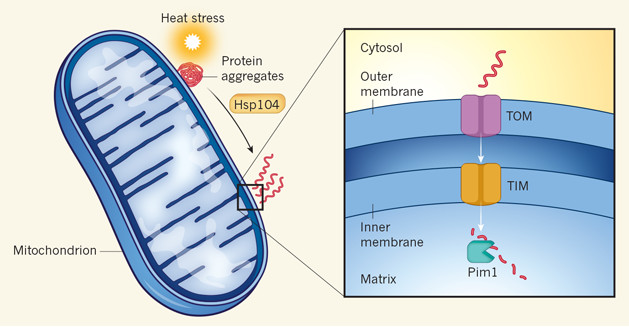
As if powering the cell wasn’t enough, mitochondria may also shoulder a share of its trash disposal duties. In the March 1 Nature, researchers led by Rong Li at Johns Hopkins University in Baltimore reported that in yeast, clumps of aggregated proteins congregate at the outer mitochondrial membrane, where a chaperone likely disentangles them before import channels funnel them into the mitochondrial matrix. There, the cytosolic riffraff meet their demise at the hands of matrix proteases. Dubbed MAGIC (mitochondria as guardian in cytosol), the process also functions in human cells, the authors claim. If extended to neurons, it could provide a tantalizing connection between two key pathologies in neurodegenerative disease: mitochondrial dysfunction and the accumulation of aggregated proteins.
“The beauty of this study is that it provides fundamental new information on how cells deal with intracellular protein aggregations,” commented Russell Swerdlow of the University of Kansas Medical Center in Kansas City. He added that the findings may help explain why researchers have spotted aggregation-prone proteins, including Aβ, in the mitochondria, and support the idea that their accumulation could interfere with mitochondrial function in neurodegenerative disease.

Munching Mitos. Protein aggregates congregate on the mitochondrial surface, where Hsp104 untangles them prior to their journey into the mitochondrial matrix for degradation. [Image courtesy of Chacinska, Nature N&V, 2017.]
Mitochondria’s claim to fame is the electron transport chain—the game of electron hot potato that produces adenosine triphosphate (ATP). But a few years ago, Li and colleagues observed yeast mitochondria doing something else: The organelles appeared to hoard aggregates on their surfaces. This held back the proteins from passing into budding daughter cells during asymmetric cell division (see Zhou et al., 2014). The clusters eventually dissolved, but not when the researchers disrupted the mitochondrial membrane potential using the toxin CCCP. This suggested that getting rid of the aggregates required fully functioning mitochondria.
For the current study, co-first authors Linhao Ruan and Chuankai Zhou and colleagues took a closer look into mitochondria’s relationship with protein clumps. First, they asked which proteins tend to aggregate during heat shock, a classic condition of stress in yeast. Using cells expressing FlucSM—a variant of luciferase that aggregates in response to heat shock and serves as a magnet for other misfolded miscreants— the researchers purified associated proteins and identified them via mass spectroscopy. Among them were components of the proteasome, chaperones, RNA binding proteins, stress granules, and elements of the mitochondrial import machinery—including Tomm40 and Tomm70.
Because of the involvement of the outer mitochondrial membrane transporters, the researchers hypothesized that aggregates might have to enter the organelles to be degraded. In support of this, Ruan found that FlucSM persisted in cells lacking functional Tim23, an import channel in the inner mitochondrial membrane.
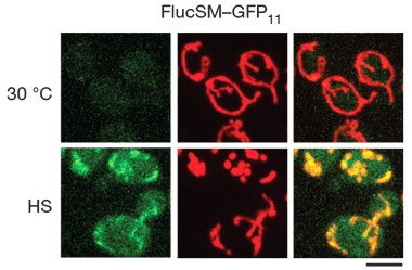
To see if tangled proteins did travel into the mitochondrial matrix, the researchers employed a split green fluorescent protein (GFP) system, in which the first 10 of 11 β-strands of GFP are ushered into the mitochondrial matrix via a mitochondrial targeting sequence (MTS), while the last β-strand is attached to some other protein of interest. Fluorescence occurs only when all strands meet up. Using this system, the researchers confirmed that upon heat shock, several aggregation-prone proteins, including FlucSM and TDP-43, crossed into the mitochondrial matrix, whereas inherently stable ones did not. High-resolution microscopy and biochemical experiments confirmed the passage of FlucSM and other aggregates, including Lsg1 and Tma19, into the matrix in response to heat shock. Hsp104, a cytosolic chaperone and disaggregator, was necessary for the translocation of FlucSM into the matrix. Li hypothesized that Hsp104 detangles protein “hairballs” on the mitochondrial surface, allowing them to squeeze through import channels.

Fluorescent MAGIC.
A split GFP (green) reconstitutes when heat shock (bottom panels) drives a GFP component that is fused with FlucSM into the mitochondrial matrix. There, it meets the other part of GFP, which is fused with mCherry (red). [Image courtesy of Ruan et al., Nature, 2017.]
What fate awaited the cytosolic emigrants upon their arrival in the matrix? The GFP signal waned after normal temperature was restored, pointing to degradation. Strikingly, inhibiting the proteasome or vacuolar proteases had little effect on the degradation of FlucSM, whereas poisoning mitochondria with CCCP did, indicating that the mitochondrial pathway was important for ridding the cytosol of stress-induced aggregates. The researchers found that the matrix protease Pim1 was required for this degradation.
MAGIC happened not only after heat shock. GFP signals also shot up in yeast at 30 degrees if the researchers inactivated Hsp70, a crucial chaperone that keeps cytosolic inhabitants neatly folded. Under these conditions, mitochondria became fragmented and ramped up production of reactive oxygen species, indicating they were under stress. The GFP signal persisted unless researchers blocked translation, hinting at a steady stream of misfolded degenerates flooding the matrix. Furthermore, so-called “super-aggregators,” such as the RNA helicase Ded1, ended up in mitochondria regardless of heat shock, painting highly unstable cytosolic proteins as matrix regulars.
Finally, the researchers asked whether MAGIC happens in mammalian cells. Employing the split-GFP system in human retinal pigment epithelium cells, the researchers found that the amount of luciferase in the matrix correlated with the protein’s stability: a super unstable variant, Fluc-DM, flooded the matrix, while Fluc-SM and wild-type Fluc passed into the organelles at lower rates. Li told Alzforum that ongoing experiments hint the pathway may operate in neurons as well, and that proteins associated with neurodegenerative diseases may be among MAGIC’s clientele.
Li speculated that MTS-containing proteins, which made up about 20 percent of those associated with mitochondria, steer co-entangled cytosolic ones toward the organelle. In regard to neurodegeneration, Li proposed that potentially toxic proteins such as α-synuclein or TDP-43 could build up in mitochondria and tax their energy production, especially as the organelle’s function already wanes with age. This, in turn, could lead to further accumulation. The constant shuttling of mitochondria up and down neuronal axons to invigorate synapses make the organelles conveniently positioned to deal with clean-ups throughout the cell, she added.
As ongoing experiments in Li’s lab investigate MAGIC in neurons, at least one neuronal example of MAGIC may already exist. Shirley ShiDu Yan of Kansas University in Lawrence previously reported that clearance of Aβ by mitochondria in neuronal synapses reduced Aβ burden and neuroinflammation, and improved learning and memory in mAPP mice (see Fang et al., 2015). In light of the current findings, Yan said she views that Aβ degradation pathway as an example of MAGIC in neuronal cells.
Flint Beal of Weill Cornell Medical College in New York added that the findings dovetail with work in his lab suggesting Aβ mingles with mitochondria in distal synapses. Li’s finding that mitochondria undergo a stress response in the face of mounting cytosolic aggregates would also be in line with the mitochondrial damage, especially in synapses, observed in neurodegenerative disease, he said.
Leonidas Stefanis of the University of Athens Medical School in Greece was impressed by the findings. “Conceptually, this is very exciting. It challenges our view of the mitochondria as basically energy-producing organelles, and suggests that they may have a considerable and specific role in removing aggregated proteins,” he wrote. “Given the importance of protein aggregation in neurodegenerative diseases at large, and the therapeutic efforts underway to enhance endogenous clearance mechanisms to combat such protein aggregation, this discovery may provide a new therapeutic target for neurodegeneration.”
Michael Lutz of Duke University in Durham, North Carolina commented that the findings provide mechanistic support for the role of the outer membrane channel Tom40 in neurodegenerative disease, including AD. The TOMM40 and ApoE genes are neighbors in the genome, and researchers led by Lutz and the late Allen Roses previously suggested that co-inheritance of these alleles predicted disease risk and age at onset (see Nov 2009 conference news and Oct 2016 news).
Mark Cookson of the National Institutes of Health commented that certain aggregation-prone proteins known to interact with the mitochondrial import machinery, such as α-synuclein, may be prime substrates for the pathway, or potentially foul it up. “Mitochondria have had a few surprises for us over the last few years, so I certainly wouldn’t dismiss this,” he said. However, he wondered about MAGIC’s overall contribution to protein turnover in neurons, given the plethora of other disposal pathways available.
In an accompanying News & Views article, Agnieszka Chacinska of the International Institute of Molecular and Cellular Biology in Warsaw, Poland, wrote that the work is a striking example of cross-talk between the cytosol and mitochondria. “It is becoming increasingly clear that maintaining a productive dialogue between cellular compartments is a crucial task—one that we are just beginning to understand.”—Jessica Shugart
References
News Citations
- Las Vegas: AD, Risk, ApoE—Tomm40 No Tomfoolery
- Field Loses Another Pioneer with Passing of Allen Roses, 73
Paper Citations
- Zhou C, Slaughter BD, Unruh JR, Guo F, Yu Z, Mickey K, Narkar A, Ross RT, McClain M, Li R. Organelle-based aggregation and retention of damaged proteins in asymmetrically dividing cells. Cell. 2014 Oct 23;159(3):530-42. Epub 2014 Oct 16 PubMed.
- Fang D, Wang Y, Zhang Z, Du H, Yan S, Sun Q, Zhong C, Wu L, Vangavaragu JR, Yan S, Hu G, Guo L, Rabinowitz M, Glaser E, Arancio O, Sosunov AA, McKhann GM, Chen JX, Yan SS. Increased neuronal PreP activity reduces Aβ accumulation, attenuates neuroinflammation and improves mitochondrial and synaptic function in Alzheimer disease's mouse model. Hum Mol Genet. 2015 Sep 15;24(18):5198-210. Epub 2015 Jun 29 PubMed.
Further Reading
Papers
- Bose A, Beal MF. Mitochondrial dysfunction in Parkinson's disease. J Neurochem. 2016 Oct;139 Suppl 1:216-231. Epub 2016 Aug 21 PubMed.
Primary Papers
- Ruan L, Zhou C, Jin E, Kucharavy A, Zhang Y, Wen Z, Florens L, Li R. Cytosolic proteostasis through importing of misfolded proteins into mitochondria. Nature. 2017 Mar 16;543(7645):443-446. Epub 2017 Mar 1 PubMed.
Annotate
To make an annotation you must Login or Register.

Comments
University of Kansas
Loss of proteostasis occurs in neurodegenerative diseases in general, and in Alzheimer’s in particular. This phenomenon, I think, is trying to tell us something important about neurodegenerative disease mechanisms. One possibility is that proteins aggregate, and that this directly causes neurodysfunction and neurodegeneration. Another is that dysfunctional, stressed, or dying cells develop and accumulate protein aggregates. A feed forward loop in which dysfunction begets aggregation which begets more dysfunction could also occur.
The beauty of this study is that it provides fundamental new information on how cells deal with intracellular protein aggregations. Here, Ruan et al. show that mitochondria themselves play a role in maintaining intracellular proteostasis, at least in yeast, but probably in mammalian cells as well. Who would have guessed that mitochondria are central players in a chaperone-mediated process through which parts of aggregated peptides are shuttled piecemeal into mitochondria, and then degraded by mitochondrial peptidases?
I think this study has potential Alzheimer’s implications. Over the past decade multiple investigators have shown aggregable proteins, including Aβ, appear at or within the mitochondrion, raising the possibility that these proteins interfere with normal mitochondrial function. Alternatively these proteins may even potentially serve a physiologically relevant role that involves modifications of mitochondria function.
In defining this phenomenon the experimental design really only looked at the ability of aggregated proteins to access mitochondria. Its goal was not to address the question of why this phenomenon evolved in the first place. At this stage of the game this is fine. Plus, I loved the last sentence, in which the authors speculate that maybe a primary mitochondrial functional decline, such as occurs in aging, could through this phenomenon go on to have a protean impact on proteostasis. It will be interesting to see what spin-offs arise from this new observation.
Weill Cornell Medical College
This is a very well done study of broad interest to scientists interested in the link between misfolded proteins and neurodegeneration. There is a great deal of evidence that mitochondria play an important role in the pathogenesis of neurodegenerative diseases, and proteins such as amyloid and α-synuclein are found either within mitochondria or in association with mitochondria-associated ER membranes (MAMs). The present paper demonstrates that aggregation-prone proteins can enter the mitochondria, where they undergo degradation. Increased import causes mitochondrial stress, which may contribute neurodegeneration.
Columbia University
Ruan and colleagues present an impressive set of data indicating an important role for mitochondria as scavengers of aggregated proteins. They term this process MAGIC, and of course if you put a new name, especially such a tantalizing one, on a cellular process, you’re onto something potentially very importan ... MAGIC is regulated, through the influence of Hsp104, by mitochondrial import, and by mitochondrial proteases, with a major role suggested for Pim1. Conceptually, this is very exciting, as it challenges our view of the mitochondria as basically energy-producing organelles, and suggests that they may have a considerable and specific role in removing aggregated proteins, something previously associated with other cellular organelles and processes, such as macroautophagy, the proteasome, and ERAD. These findings thus touch on fundamental aspects of cell biology.
Given the importance of protein aggregation in neurodegenerative diseases at large, and the therapeutic efforts underway to enhance endogenous clearance mechanisms to combat such protein aggregation, this discovery may provide a new therapeutic target for neurodegeneration. The authors use an impressive array of cell biology and biochemical assays mainly in yeast cells. It is important that, under the admittedly somewhat artificial conditions of heat shock, MAGIC was a major pathway for aggregate clearance, compared to other established processes. However, generalization of the importance of this process to mammalian cells, in particular in an in vivo situation, remains to be demonstrated. A small set of experiments in mammalian cells does suggest, however, that MAGIC may be operating in such systems as well, but its comparative importance to other protein degradation systems and its role in endogenous aggregated protein turnover were not assessed. The exact molecular pathway of recognition and uptake of aggregated proteins into mitochondria also requires further elucidation.
Duke University School of Medicine
Mitochondrial dysfunction is increasingly being investigated as a pathophysiological contributor to neurodegenerative diseases including Alzheimer’s disease (AD). Several mitochondrial cellular processes are involved in the biogenesis, folding, trafficking, and clearance of proteins that maintain cytosolic proteostasis. This paper describes the results of biochemical studies in yeast to investigate the import of protein aggregates into mitochondria. Notably, in response to heat shock, protein aggregates form, interact with both cytosolic and mitochondrial proteins, and are imported into mitochondria. Translocase of the outer mitochondrial membrane proteins (Tomm70 and Tomm40), which transport peptides and proteins into the mitochondria, were found to co-purify with the protein aggregates, evidence for interaction. Defects in a cytosolic heat shock protein (Hsp70) were found to accelerate the import of misfolded proteins into the mitochondria and in turn increase mitochondrial stress.
To conceptualize the results, the authors develop the concept of mitochondria as guardian in cytosol (MAGIC), in other words, the mitochondria has a specific role in facilitating protein disaggregation in the cytosol by removing and transporting dissociated proteins into the mitochondria in response to stress. As a consequence, the dissociated proteins could accumulate in mitochondria and have an impact on development of disease. Genetic and biochemical data have supported a role for Tomm40 in late-onset neurodegenerative diseases including AD; the present study provides specific data on cellular interactions that may offer a mechanistic interpretation for this role. The results discussed in the paper and the approach are innovative in terms of stimulating new thinking about the underlying neurobiology of AD, specifically the involvement of mitochondria and outer membrane proteins including Tomm40.
University of Kansas
Ruan et al. reported a novel mitochondrial function in yeast in preventing protein aggregation. The authors demonstrated that heat shock stress can induce extensive protein aggregates in yeast and that normal mitochondria play an important role in eliminating these unhealthy aggregates. This process depends on HSP104, TIM23, PIM1, as well as an intact mitochondrial membrane potential. Although there is significant distinction between yeast and mammalian cell mitochondria, the mitochondria as guardian in cytosol (MAGIC) pathway will also be expected in the human tissues, besides numerous recently discovered new functions for mitochondria.
Further studies to identify similar pathways in mammalian cells or neurons will be important steps in discovering therapy for neurodegenerative disorders. Our previous work had shown an important role of mitochondrial proteases in processing amyloid peptide generated in the cytosol, which can be an example of MAGIC pathway in neuronal cells. Presequenceprotease (PreP), a mitochondrial peptidasome, is a novel mitochondrial Aβ degrading enzyme. We have demonstrated that PreP was significantly reduced in AD brains and in transgenic Alzheimer’s disease mouse models that overexpress human Aβ (Alikhani et al., 2011). Importantly, increased neuronal PreP activity attenuated cerebral and mitochondrial pools of Aβ, and rescued mitochondrial and synaptic function, as well as learning and memory (Fang et al., 2015). These results suggest that PreP functions as a peptide scavenger, clearing mitochondria of Aβ, and thereby protecting mitochondria against pathogenic peptide intruders. We also unexpectedly observed that increased expression of PreP not only degrades mitochondrial Aβ but also affects total brain Aβ levels, suggesting that mitochondrial Aβ is not just a “spilling over” from cellular aggregation and that PreP also has an important regulating effect on total brain Aβ levels. Given that exogenous or intracellular Aβ is capable of direct transport into mitochondria via mitochondrial channel proteins such as TOMM40, the receptor for advanced glycation end product (RAGE), or by an unknown mechanism, the mitochondrial pool of Aβ may undergo dynamic changes in different intracellular compartments, contributing to the balance of intracellular/extracellular Aβ accumulation.
References:
Alikhani N, Guo L, Yan S, Du H, Pinho CM, Chen JX, Glaser E, Yan SS. Decreased proteolytic activity of the mitochondrial amyloid-β degrading enzyme, PreP peptidasome, in Alzheimer's disease brain mitochondria. J Alzheimers Dis. 2011 Jan 1;27(1):75-87. PubMed.
Fang D, Wang Y, Zhang Z, Du H, Yan S, Sun Q, Zhong C, Wu L, Vangavaragu JR, Yan S, Hu G, Guo L, Rabinowitz M, Glaser E, Arancio O, Sosunov AA, McKhann GM, Chen JX, Yan SS. Increased neuronal PreP activity reduces Aβ accumulation, attenuates neuroinflammation and improves mitochondrial and synaptic function in Alzheimer disease's mouse model. Hum Mol Genet. 2015 Sep 15;24(18):5198-210. Epub 2015 Jun 29 PubMed.
Make a Comment
To make a comment you must login or register.