Ring Around the Vessel: Enlarged Spaces Signal Vascular Disease
Quick Links
In the July 17 JAMA Neurology, scientists led by Lenore Launer, National Institute on Aging, Bethesda, Maryland, report that enlarged spaces around the brain’s blood vessels are associated not only with cerebrovascular trouble but also with cognitive decline and vascular dementia. Visible in magnetic resonance imaging (MRI), these gaps are called large perivascular spaces (L-PVSs). They could represent a new marker that heralds cerebrovascular disease and vascular dementia.
“This prospective study demonstrates that L-PVSs are associated with small vessel disease and development of vascular dementia,” said Roxana Carare, University of Southampton, England, in the U.K.
“This is a relatively new marker,” Launer told Alzforum. “We would like to see it incorporated into a risk score, where several markers of small vessel disease would be a robust predictor of whether someone is at increased risk for dementia.”

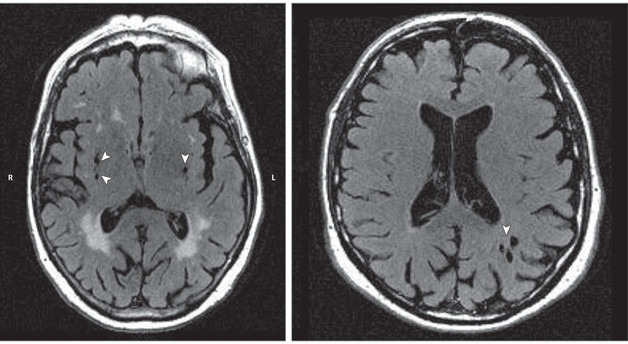
Holes Herald Trouble. Large perivascular spaces (white arrowheads) appear around blood vessels in the basal ganglia (left) and white matter (right). [© 2017, American Medical Association. All rights reserved.]
The areas surrounding blood vessels are normally microscopic. They help drain fluid and metabolic waste from the brain. However, these spaces expand with age and some become visible by MRI, appearing as round or tubular hyperintensities on scans.
Epidemiologic data suggest that PVSs go hand in hand with small vessel disease and cognitive impairment (Ramirez et al., 2015; Zhu et al., 2010). However, longitudinal data examining the association is scarce (Zhu et al., 2010). What’s more, most studies focus on small PVSs under 3 mm in diameter. Launer’s team wanted to test the link prospectively, and focus on PVSs bigger than 3 mm, considered a more severe form of the lesion.
To do this, first author Jie Ding and colleagues used data from the prospective Age, Gene/Environment Susceptibility–Reykjavik (AGES-Reykjavik) Study. Sampling from the population, 5,764 people had a baseline MRI and cognitive testing between 2002 and 2006. The tests measured verbal memory, processing speed, and executive function. Of the original sample, 2,612 returned for both follow-up MRI and cognitive tests between 2007 and 2011. The researchers measured white-matter hyperintensities, microbleeds, and subcortical infarcts—all signs of small vessel disease.
At the five-year mark, 424 people had L-PVSs. At baseline, people with L-PVSs had more subcortical infarcts, microbleeds, and white-matter hyperintensity progression than people without L-PVSs. The more a person had, the more advanced their small vessel disease.
The cognitive function measurably associated with L-PVSs over the five-year follow-up was processing speed. It slowed in people with L-PVSs, most dramatically in those who had two or more of the markers at baseline. They were also four times more likely to develop clinical vascular dementia, diagnosed according to the criteria of the State of California Alzheimer Disease Diagnostic and Treatment Centers (Chui et al., 1992). However, their risk for all-cause dementia or Alzheimer’s disease stayed the same as that of the general population, meaning L-PVSs constitute a biomarker for vascular, but not other types of dementia.
The researchers remain unsure whether L-PVSs or small vessel disease comes first. Baseline L-PVSs were often seen at the same time as markers of small vessel disease, so it’s not clear. It’s also unclear how strong the association truly is. The scientists cautioned that people who stayed on for follow-up tended to be younger, more educated, and healthier at baseline than those who dropped out. In addition, those who had more severe vascular disease died or were lost to follow-up. That means the association may have gone underestimated.
“This interesting observation adds one more potential biomarker for vascular dementia,” said Costantino Iadecola, Weill Cornell Medical College, New York. “It will probably contribute to a constellation of imaging findings that will speak to an increased incidence of cognitive impairment or vascular disease of the brain.” L-PVSs could help explain why two people with otherwise equal cerebrovascular loads could be cognitively affected to different degrees, he said.
At L-PVSs, clearance slows down and solutes can build up. “Targeting the failure of clearance of fluid along these drainage pathways could be an efficient early therapeutic strategy for aging brains in the prevention of vascular cognitive impairment,” Carare said. However, this will be hard to pull off until researchers clarify the exact mechanism behind the L-PVSs (and its impact on the clearance process), Iadecola added. —Gwyneth Dickey Zakaib
References
Paper Citations
- Ramirez J, Berezuk C, McNeely AA, Scott CJ, Gao F, Black SE. Visible Virchow-Robin spaces on magnetic resonance imaging of Alzheimer's disease patients and normal elderly from the Sunnybrook Dementia Study. J Alzheimers Dis. 2015;43(2):415-24. PubMed.
- Zhu YC, Tzourio C, Soumaré A, Mazoyer B, Dufouil C, Chabriat H. Severity of dilated Virchow-Robin spaces is associated with age, blood pressure, and MRI markers of small vessel disease: a population-based study. Stroke. 2010 Nov;41(11):2483-90. Epub 2010 Sep 23 PubMed.
- Zhu YC, Dufouil C, Soumaré A, Mazoyer B, Chabriat H, Tzourio C. High degree of dilated Virchow-Robin spaces on MRI is associated with increased risk of dementia. J Alzheimers Dis. 2010;22(2):663-72. PubMed.
- Chui HC, Victoroff JI, Margolin D, Jagust W, Shankle R, Katzman R. Criteria for the diagnosis of ischemic vascular dementia proposed by the State of California Alzheimer's Disease Diagnostic and Treatment Centers. Neurology. 1992 Mar;42(3 Pt 1):473-80. PubMed.
Further Reading
Papers
- Banerjee G, Kim HJ, Fox Z, Jäger HR, Wilson D, Charidimou A, Na HK, Na DL, Seo SW, Werring DJ. MRI-visible perivascular space location is associated with Alzheimer's disease independently of amyloid burden. Brain. 2017 Apr 1;140(4):1107-1116. PubMed.
- Ramirez J, Berezuk C, McNeely AA, Gao F, McLaurin J, Black SE. Imaging the Perivascular Space as a Potential Biomarker of Neurovascular and Neurodegenerative Diseases. Cell Mol Neurobiol. 2016 Mar;36(2):289-99. Epub 2016 Mar 18 PubMed.
Primary Papers
- Ding J, Sigurðsson S, Jónsson PV, Eiriksdottir G, Charidimou A, Lopez OL, van Buchem MA, Guðnason V, Launer LJ. Large Perivascular Spaces Visible on Magnetic Resonance Imaging, Cerebral Small Vessel Disease Progression, and Risk of Dementia: The Age, Gene/Environment Susceptibility-Reykjavik Study. JAMA Neurol. 2017 Sep 1;74(9):1105-1112. PubMed.
Annotate
To make an annotation you must Login or Register.

Comments
University of Southampton School of Medicine
In this study, Ding et al. followed up MRI and cognitive changes in 2,612 patients enrolled in the population-based Age, Gene/Environment Susceptibility–Reykjavik study over five years, with particular attention to large perivascular spaces over 3 mm in diameter. This prospective study demonstrates that LPVS are associated with small vessel disease and development of vascular dementia.
LPVS are present in the white matter or basal ganglia, where the periarterial compartment is different compared to the cortical grey matter. A layer of leptomeninges reflects over an artery as it enters the cortex from the subarachnoid space. This layer closely adheres to the wall of the artery and, due to the tight extracellular spaces, there is no potential for accumulation of fluid.
There are two such layers in the walls of arteries of the white matter and basal ganglia, with potential for enlargement due to excess fluid, most likely observed on MRI as LPVS. The authors recognize the possibility that this enlargement occurs as a result of a failure of drainage of solutes and fluid along the intramural periarterial drainage pathways. Targeting the failure of clearance of fluid along the intramural periarterial drainage pathways could be an efficient early therapeutic strategy for the aging brain in the prevention of vascular cognitive impairment.
Johns Hopkins University School of Medicine
This study adds to our expanding knowledge about the importance of enlarged perivascular spaces regarding brain outcomes. It used to be thought that these perivascular spaces were not pathologic at all, but recent studies have emphasized that they share similar risk factors (e.g., hypertension) to other markers of small vessel disease in the brain, such as white-matter hyperintensities or lacunar infarcts or microbleeds.
This study in the AGES-Reykjavik cohort further emphasizes the importance of perivascular spaces as a marker of not only other forms of small vessel disease, but also of poor cognitive outcomes. It is unclear, however, whether the spaces themselves are meaningful for cognitive outcomes, or are simply markers of the overall burden of small vessel disease. Even though the associations in this study were independent of those other small vessel markers, it’s likely that we cannot visualize all forms of small vessel disease on current MRI scans.
This means that we may not necessarily focus future treatments on reducing large perivascular spaces, but they may be important biomarkers for small vessel disease more broadly, which is something we’d like to prevent and treat.
Sunnybrook Research Institute, University of Toronto
Sunnybrook Health Sciences Centre, University of Toronto
This article is a novel contribution, which looks at how MRI-visible perivascular spaces (PVS) are associated with progression of small vessel disease and dementia risk.
The results from this Icelandic study are interesting as it specifically examines large PVS. Previous consensus criteria, such as the STRIVE criteria, defined PVS as being smaller than 3mm in diameter (with the acknowledgement that there are some larger PVS in rare cases). Our group at the Sunnybrook Research Institute at the University of Toronto and others have periodically observed PVS in Alzheimer’s disease patients that exceed the 3mm STRIVE criteria, mostly in the basal ganglia. The findings from Ding and colleagues examining an impressive 2,612 participants confirm such anecdotal observations, reporting a prevalence of 16 percent, with the majority of them occurring in the basal ganglia (82 percent).
The findings in this study demonstrating associations of large PVS with other markers of small vessel disease, dementia risk, and information processing speed, may suggest that the consensus-based criteria should be revised to include PVS exceeding the 3mm diameter definition. However, as acknowledged by the co-authors in their limitations, this may be more difficult than it seems, as in many cases lacunar infarcts have similar appearance on MRI and many do not present with a hyperintense rim on FLAIR. The less popular proton density MRI may be prove useful in future studies as PVS are not hyperintense on PD.
Interestingly, the findings of this study are in contrast to the recently published findings from the large (n=1,228) Northern Manhattan Study (Guttierrez et al., 2017), which demonstrated that a higher burden of small PVS (≤3mm), as opposed to large, were associated with vascular events such as stroke, myocardial infarction, and death.
Large versus small, small number of large versus high number of small—which is worse remains unclear. As Ding and colleagues state, it is likely that these issues arising from PVS rating scales may be resolved with future implementation of quantitative methods for measuring PVS. Such methods are currently being developed by our lab in collaboration with Drs. Wardlaw, Zlokovic, Nedergaard, Charpak, Benveniste, and Smith, as part of a Leducq Fondation grant examining the role of PVS in small vessel disease utilizing experimental and translational clinical approaches.
The recent explosion of research examining PVS is particularly relevant to Alzheimer’s disease and dementia, since it has implications for the glymphatic system—a complex series of perivascular channels that the brain uses to clear waste, such as amyloid, out of the brain. However, as we have outlined, there remain many challenges to overcome before MRI-visible PVS can be utilized as a target for prevention/treatment in Alzheimer’s and related disorders.
Our work was featured in a previous article on Alzforum regarding sleep disturbances, PVS, and clearance (see May 2016 news).
References:
Gutierrez J, Elkind MS, Dong C, Di Tullio M, Rundek T, Sacco RL, Wright CB. Brain Perivascular Spaces as Biomarkers of Vascular Risk: Results from the Northern Manhattan Study. AJNR Am J Neuroradiol. 2017 May;38(5):862-867. Epub 2017 Mar 24 PubMed.
University of Edinburgh
University of Southern California
University of Southern California
There is increasing evidence that enlarged perivascular spaces (PVSs) are associated with cerebrovascular dysfunction, as well as with cognitive decline and dementia. Using combined T2-weighted fluid attenuated inversion recovery (FLAIR) and T2*-weighted gradient-echo echo-planar imaging (GRE-EPI) magnetic resonance imaging (MRI) sequences, Ding and colleagues studied large PVSs (LPVSs)—bigger than 3 mm diameter—in 2,612 individuals. All individuals enrolled in the study had MRI and cognitive testing at baseline and after five years follow-up. Individuals with dementia at baseline were excluded from the study.
Out of the 2,612 participants, 424 (16 percent) had LPVSs at baseline. After adjusting for age, sex, and interval between baseline and follow-up scanning, at a five-year follow-up individuals with LPVSs at baseline compared to those without LPVSs developed imaging biomarkers of small vessel disease (SVD) including white-matter hyperintensities (WMH), subcortical infarcts, and microbleeds. Moreover, there was a correlation between the number of LPVSs and decline in the processing speed—measured with Digit Symbol Substitution test (DSST), the Figure Comparison Test, and the Stroop Test Part I (word naming) and Part II (color naming)—after five years. No association was found with verbal memory and executive function.
Interestingly, the authors suggested that the risk for Alzheimer’s disease (AD) did not change in participants with or without LPVSs at baseline. However, whether LPVSs can contribute to pathophysiology of AD still remains unclear in our opinion, as Alzheimer’s imaging or molecular biomarkers such as amyloid and tau load and/or Aβ and tau cerebrospinal fluid levels, have not been determined in this study. It is also unclear whether LPVSs precede imaging biomarkers of SVD or not, as some participants having at baseline LPVSs also had SVD biomarkers.
LPVSs have been speculated to be an imaging biomarker for early vascular dysfunction and possibly blood-brain barrier (BBB) breakdown that may eventually lead to the entry and perivascular brain accumulation of blood-derived neurotoxic molecules (e.g., fibrinogen, plasminogen, and thrombin, among others), cells (e.g., red blood cells, leucocytes), and fluid, and even sometimes microbial pathogens (Zhao et al., 2015).
BBB breakdown, on the other hand, has been established in different types of dementia including vascular, AD, and other neurodegenerative disorders. Therefore, it would be helpful for future studies to determine whether BBB breakdown is related to LPVSs and/or SVD. Indeed, we recently found an age-dependent BBB breakdown in cognitively normal individuals, and worsening of BBB disruption in early stages of cognitive impairment, as demonstrated by dynamic contrast-enhanced (DCE)-MRI method (Montagne et al., 2015). A longitudinal study including T2w FLAIR/T2*w GRE-EPI sequences for LPVSs measurement, T2*-weighted for hemosiderin deposits, and DCE-MRI for BBB permeability might shed more light on the early vascular events occurring in SVD patients.
References:
Montagne A, Barnes SR, Sweeney MD, Halliday MR, Sagare AP, Zhao Z, Toga AW, Jacobs RE, Liu CY, Amezcua L, Harrington MG, Chui HC, Law M, Zlokovic BV. Blood-brain barrier breakdown in the aging human hippocampus. Neuron. 2015 Jan 21;85(2):296-302. PubMed.
Zhao Z, Nelson AR, Betsholtz C, Zlokovic BV. Establishment and Dysfunction of the Blood-Brain Barrier. Cell. 2015 Nov 19;163(5):1064-78. PubMed.
Make a Comment
To make a comment you must login or register.