Some Neurons Enlist Glia to Take Care of Their Trash
Quick Links
Normally, cells recycle mitochondria by breaking them down in lysosomes in a process called mitophagy. However, new work suggests that certain neurons delegate the cleanup to nearby glia. Scientists led by Nicholas Marsh-Armstrong, The Johns Hopkins University School of Medicine, Baltimore, and Mark Ellisman, University of California, San Diego, found that the axons of retinal ganglion cells expel bundles containing mitochondria into the surrounding milieu. Neighboring astrocytes then appear to gobble them up.
“This is a very interesting discovery, as it describes a completely novel process through which neurons can get rid of their mitochondria,” Ben Barres, Stanford University School of Medicine, California, wrote to Alzforum in an email. Barres was not involved in the study, which appeared online June 16 in the Proceedings of the National Academy of Sciences.

Blowing out the Trash
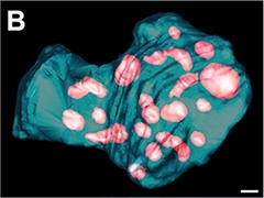
Axons eject bundles (turquoise) containing mitochondria (pink) that are later broken down by astrocytes. Courtesy of Davis et al. 2014. PNAS.
Previously, Marsh-Armstrong and colleagues noticed protrusions sprouting from the axons of healthy retinal ganglion cells in the optic nerve head (ONH) of normal mice. These were engulfed by astrocytes (see Nguyen et al., 2011). In the current study, they used electron microscopy to see what was inside these bundles.
In two mice, one three months old and the other nine months old, the clumps contained full and fragmented mitochondria, complete with their characteristic inner membrane folds, or cristae. Each package contained about 30 mitochondria. The axons that released them seemed healthy. The researchers also found microtubules and unidentified flotsam inside the bundles, suggesting the axons disposed of additional components this way.
To confirm the organelles came from neurons, first author Chung-ha Davis and colleagues introduced a fluorescent mitochondrial reporter into the retinal neurons of 29 mice. Many labeled mitochondria ended up in astrocytes at the optic nerve head. Almost five times more mitochondria were broken down there than were degraded inside neuron bodies.
Given that the optic nerve head is a specialized structure, the researchers wondered whether this phenomenon, which they termed “transmitophagy,” occurred elsewhere. When they looked near serotonergic neurons of the cerebral cortex, they found some evidence of it, hinting that transmitophagy could occur in other areas of the nervous system, the authors wrote.
While it’s unclear why neurons might use transmitophagy, the researchers speculate that it helps long axons eliminate garbage by giving it a more efficient disposal system than transporting it all back to the soma, where the neuronal lysosomes reside. Neurons overwhelmed by their mitochondrial load or under stress might also rely on this mechanism, they suggested. This expulsion of axon components could explain how material typically found inside neurons ends up outside, for example proteins linked to neurodegenerative disease, such as Aβ and α-synuclein, Marsh-Armstrong told Alzforum. He speculated that dysfunctional glial processing could contribute to extracellular build-up of toxic proteins. He said his group next plans to check whether this process malfunctions in animal models of glaucoma. This eye disease causes loss of retinal ganglion cells and scientists hypothesize that mitochondrial dysfunction may be involved (see Kong et al., 2009). The researchers will also check to see where else this process takes place in the central nervous system.
Outside experts were intrigued by the finding (see comments below). “It’s a fundamental piece of physiology that wasn’t generally recognized before,” said Russell Swerdlow, University of Kansas, Kansas City. He noted that the study could have implications for neuroinflammation. Mitochondria retain some of their prokaryotic roots, he explained, so if a breakdown in transmitophagy abandons mitochondria outside the cell, the immune system might interpret them as foreign and mount a response. In the big picture, the results contribute to the evolving notion that astrocytes have important intricate physiological relationships with neurons, he said.—Gwyneth Dickey Zakaib
References
Paper Citations
- Nguyen JV, Soto I, Kim KY, Bushong EA, Oglesby E, Valiente-Soriano FJ, Yang Z, Davis CH, Bedont JL, Son JL, Wei JO, Buchman VL, Zack DJ, Vidal-Sanz M, Ellisman MH, Marsh-Armstrong N. Myelination transition zone astrocytes are constitutively phagocytic and have synuclein dependent reactivity in glaucoma. Proc Natl Acad Sci U S A. 2011 Jan 18;108(3):1176-81. PubMed.
- Kong GY, Van Bergen NJ, Trounce IA, Crowston JG. Mitochondrial dysfunction and glaucoma. J Glaucoma. 2009 Feb;18(2):93-100. PubMed.
Further Reading
Primary Papers
- Davis CH, Kim KY, Bushong EA, Mills EA, Boassa D, Shih T, Kinebuchi M, Phan S, Zhou Y, Bihlmeyer NA, Nguyen JV, Jin Y, Ellisman MH, Marsh-Armstrong N. Transcellular degradation of axonal mitochondria. Proc Natl Acad Sci U S A. 2014 Jul 1;111(26):9633-8. Epub 2014 Jun 16 PubMed.
Annotate
To make an annotation you must Login or Register.

Comments
University of Southern California
Davis and coworkers have uncovered an exciting neurobiological phenomenon whereby mitochondria in retinal ganglion cells are "shed" and taken up by nearby astrocytes. The authors are dubbing this "transmitophagy," or transcellular degradation of mitochondria. As duly pointed out by the authors, this exciting new finding likely occurs in other CNS regions. While the authors have focused in this report on physiological transmitophagy, their results nonetheless beg the question of what happens vis-a-vis pathological CNS states like Alzheimer’s disease.
University of Kansas
This is an exciting new finding on a pathway of neuronal mitochondrial degradation and clearance. The authors provide convincing data showing a process through which the axonal mitochondria can be shed through evulsion and then be phagocytosed by adjacent astrocytes. This is a new pathway for maintenance of mitochondrial health in addition to mitochondrial fission, fusion, and mitophagy, and it is possibly unique for neuronal axons. This phenomenon is found in a healthy mouse optic nerve head after intravitreal injection of a reporter gene. The optic nerve head is a region crowded with axons without myelination and it is very vulnerable to increased tissue pressure. Therefore, it is not clear whether this is a normal phenomenon or this is related to early axonal injury. Interestingly, “transmitophagy” was also found in the cerebral cortex, although much less frequently. There is a possibility that microglia besides astrocytes might also play a role in clearance of unhealthy mitochondria within the nervous system.
Further study is necessary to elucidate the signaling and cellular pathways involved in the transcellular mitophagy, which might help us understand how axons maintain their health through restoration of mitochondrial integrity and the pathogenesis of various neurological disorders such as neurodegenerative disorders.
I think this is a very interesting discovery as it describes a completely novel process, transmitophagy, through which neurons can get rid of their mitochondria. Recent studies, including our own, have indicated that astrocytes are highly phagocytic cells, so it will be interesting to see if specific astrocyte phagocytic pathways are mediating this transmitophagy. It will also be extremely interesting to know if defects in this process could lead to neurodegenerative disease. For instance, when the retinal pigment cells, which are phagocytic, are defective in their ability to eat the "shed" used up portions of photoreceptor outer segments, a retinal degenerative disorder results.
This is truly an exciting paper with an elegant demonstration of extrusion of mitochondria and potentially other axonal components into the extracellular milieu and reuptake by glia. This process, named transmitophagy by the authors, has also been described previously by three-dimensional electron microscopy in aged rhesus monkeys (Fiala et al., 2007) and the hippocampi of aged wild-type mice (Doehner et al., 2012).
Interestingly, in wild-type mice that were prenatally exposed to a viral–like challenge that resulted in AD-like changes in aged animals (Krstic et al., 2012), the number of these transmitophagy events increased, and each consisted of higher mitochondria density (Doehner et al., 2012).
In line with the Davis et al.’s hypothesis that transmitophagy may be an efficient garbage disposal system of long-projection neurons, we proposed recently that brain injury (e.g., brain and axonal damage, chronic neuroinflammation, oxidative stress, etc.) might impair the transmitophagy process and result in enlargement and concomitant disruption of these axonal buddings (Krstic and Knuesel, 2013). This phenomenon was, in fact, described in a postmortem tracing study in human AD brain slices (Xiao et al., 2011).
Altogether, the findings presented here provide a first functional insight into a putative protective axonal mechanism that has been described earlier and has served as basis for a novel cellular mechanism of senile plaque and neurofibrillary tangle formation in late-onset Alzheimer’s disease. (Krstic and Knuesel, 2013).
One could go even further and speculate that the fibrillary amyloid plaques might actually represent a hydrophobic seal of a source of ECM-floating mitochondria and hence be an aide de camp in the neuronal battle against neurodegeneration. It would not be surprising, therefore, to find a regulatory role of APP in the transmitophagy process. (Krstic and Knuesel, 2013).
References:
Fiala JC, Feinberg M, Peters A, Barbas H. Mitochondrial degeneration in dystrophic neurites of senile plaques may lead to extracellular deposition of fine filaments. Brain Struct Funct. 2007 Sep;212(2):195-207. Epub 2007 Aug 17 PubMed.
Doehner J, Genoud C, Imhof C, Krstic D, Knuesel I. Extrusion of misfolded and aggregated proteins--a protective strategy of aging neurons?. Eur J Neurosci. 2012 Jun;35(12):1938-50. PubMed.
Krstic D, Madhusudan A, Doehner J, Vogel P, Notter T, Imhof C, Manalastas A, Hilfiker M, Pfister S, Schwerdel C, Riether C, Meyer U, Knuesel I. Systemic immune challenges trigger and drive Alzheimer-like neuropathology in mice. J Neuroinflammation. 2012;9:151. PubMed.
Krstic D, Knuesel I. Deciphering the mechanism underlying late-onset Alzheimer disease. Nat Rev Neurol. 2013 Jan;9(1):25-34. PubMed.
Xiao AW, He J, Wang Q, Luo Y, Sun Y, Zhou YP, Guan Y, Lucassen PJ, Dai JP. The origin and development of plaques and phosphorylated tau are associated with axonopathy in Alzheimer's disease. Neurosci Bull. 2011 Oct;27(5):287-99. PubMed.
Krstic D, Knuesel I. The airbag problem-a potential culprit for bench-to-bedside translational efforts: relevance for Alzheimer's disease. Acta Neuropathol Commun. 2013 Sep 23;1(1):62. PubMed.
Make a Comment
To make a comment you must login or register.