MRI Scans Reveal ALS Features in Upper Motor Neurons
Quick Links
Magnetic resonance imaging detects upper motor neuron damage in people with amyotrophic lateral sclerosis, and the signal correlates with the severity of a patient’s symptoms, according to a small study published in the August PLoS One.
Scientists at the Perelman School of Medicine at the University of Pennsylvania in Philadelphia scanned 34 people with ALS using a mode called diffusion tensor imaging (DTI) to investigate how water molecules “wiggle about” in brain tissues, explained senior author John Woo. DTI allows researchers to visualize white-matter structures such as the corticospinal tract, where upper motor neurons reside. Woo and co-authors improved on previous DTI studies by using unbiased, computational analyses to measure increased water diffusion in this tract. This movement reflects degradation of neurons, scientists suspect, because healthy, space-filling axon tracts restrict water flow. DTI measurements might make a useful biomarker for ALS, Woo suggested, though he emphasized that more analysis is required to understand those structural changes and apply them in trials or in the clinic.
Use of DTI in neurodegenerative diseases generally has been on the rise over the past several years as scanners and processing algorithms improved (e.g., Abhinav et al., 2014). Two measures that quantify water movement are mean diffusivity and fractional anisotropy. Mean diffusivity (MD), Woo explained, describes how far water molecules can move in the tissues. The farther they travel, the higher the MD. Fractional anisotropy (FA) considers not just distance but direction. Solid structures interfere with water’s ability to move in some directions more than others, and higher FA values reflect this restricted diffusion. For example, water moves more easily along an axon’s length than it does across the axon, so that structure would have a high FA.
Scientists have been studying ALS with DTI over the last two decades. In 1999, people with the disease were found to have higher MD and lower FA in the corticospinal tract, where upper motor neurons reside (Ellis et al., 1999). Reduced FA correlated with more severe upper motor neuron symptoms and MD increased with longer disease duration.

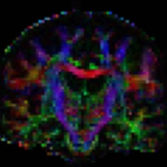
Diffusion tensor images indicate how freely water molecules move in the front-to-back (blue), up-down (green), and left-right (red) dimensions. [Image courtesy of Woo et al., 2014.]
Since then, the correlation between disease and DTI measurements has been controversial. Some studies confirmed the high-MD, low-FA measurements for ALS (Cosottini et al., 2005; Wang et al., 2006; Sage et al., 2007). However, others were unable to replicate the correlation with clinical symptoms and progression that Ellis et al. observed (Toosy et al., 2003; Ciccarelli et al., 2006). Woo suggested the discrepancies may result from disparate DTI methods. In early studies, researchers outlined the corticospinal tract by hand, which could introduce error. In later work, some authors used different kinds of computer algorithms to delineate the tract. The MD and FA values will change depending on how the corticospinal tract is defined, and Woo suggested this kind of variation could explain the discrepancies.
In the PLoS One study, the researchers applied a new method to standardize DTI processing. One challenge with neural imaging is that people have differently sized and shaped brains, making it hard to compare them. To address this, Woo and colleagues created an “average” brain shape from their 34 participants. They then used computer software, rather than manual tracing, to outline the corticospinal tract in that average image. The researchers used this average as a template, morphing each individual brain image to fit. Individual corticospinal tracts had the same size and shape as the template, but retained their unique water-diffusion characteristics. Therefore, the scientists could calculate MD and FA in all 34 scans in the same way, eliminating some of the variability that plagued previous studies. “This is a very powerful way of [analyzing DTI] in an unbiased fashion,” commented Erik Pioro of the Cleveland Clinic, who was not involved in the current work.
Another improvement in Woo’s methods was in a new clinical scale the researchers used to assess disease, focusing specifically on upper motor neurons. Neurologists Leo McCluskey and Lauren Elman, study co-authors, assigned points for specific upper motor neuron-based symptoms in the head and limbs, such as abnormal reflexes. They dubbed this number, which ranged from 0 to 32, the Penn UMN score. Pioro praised the scale, calling the parameters “well thought-out.”
With the improved image handling, Woo and colleagues confirmed Ellis et al.’s 1999 findings. Compared with 13 control subjects with no neurologic disease, people with ALS had higher MD and lower FA. Also, among the people with ALS, there was a linear relationship between the DTI measures and the Penn UMN score. Those with the more severe symptoms tended to have raised MD and reduced FA.
“I would like to think of DTI as a promising method to noninvasively study the brain in ALS,” Woo said. For now, he cannot be certain how DTI might be applied as a biomarker, for example as a diagnostic or prognostic test. Several challenges remain to develop the technique for widespread use. For one, imaging protocols vary between sites. “Even replicating DTI results from one scanner to the next is fraught with difficulty,” Woo said. In the Alzheimer’s field, scientists have begun to address this standardization issue with the large-scale Alzheimer’s Disease Neuroimaging Initiative (see Oct 2008 news story), and continue to discuss how to delineate brain structures, such as the hippocampus, in a standardized way (see Aug 2012 conference story). The ALS field may need a similar approach, Woo said.
Another difficulty with DTI is that researchers are not sure precisely what they are imaging. “We still do not really understand what it represents pathologically,” Pioro said. He is conducting an analysis of DTI and postmortem features.
It also will be important, Pioro said, to conduct longitudinal studies of DTI in ALS. “Can we detect progression of these abnormalities that we can then use as a readout for a clinical trial?” he wondered. Woo expressed an interest in that as well, but longitudinal studies are difficult with people who have ALS. Because the disease frequently takes months to diagnose, people have already passed the earliest stages once they are ready to participate in a trial. In the latter stages of disease, people have trouble lying in an MRI scanner.
Woo wants to examine different segments of the corticospinal tract and compare the water diffusion there to the location of people’s motor symptoms. For example, he predicts that a person who can’t move his or her left leg would exhibit altered DTI measures in the upper motor neurons that control that limb. This kind of analysis will require slightly different imaging techniques than used in the current study, he said (Graham et al., 2004; Rose et al., 2012).—Amber Dance
References
News Citations
- ADNI Results: A Story of Standardization and Science
- Can We All Agree on How to Draw a Hippo(campus)?
Paper Citations
- Abhinav K, Yeh FC, Pathak S, Suski V, Lacomis D, Friedlander RM, Fernandez-Miranda JC. Advanced diffusion MRI fiber tracking in neurosurgical and neurodegenerative disorders and neuroanatomical studies: A review. Biochim Biophys Acta. 2014 Nov;1842(11):2286-2297. Epub 2014 Aug 12 PubMed.
- Ellis CM, Simmons A, Jones DK, Bland J, Dawson JM, Horsfield MA, Williams SC, Leigh PN. Diffusion tensor MRI assesses corticospinal tract damage in ALS. Neurology. 1999 Sep 22;53(5):1051-8. PubMed.
- Cosottini M, Giannelli M, Siciliano G, Lazzarotti G, Michelassi MC, Del Corona A, Bartolozzi C, Murri L. Diffusion-tensor MR imaging of corticospinal tract in amyotrophic lateral sclerosis and progressive muscular atrophy. Radiology. 2005 Oct;237(1):258-64. PubMed.
- Wang S, Poptani H, Woo JH, Desiderio LM, Elman LB, McCluskey LF, Krejza J, Melhem ER. Amyotrophic lateral sclerosis: diffusion-tensor and chemical shift MR imaging at 3.0 T. Radiology. 2006 Jun;239(3):831-8. Epub 2006 Apr 26 PubMed.
- Sage CA, Peeters RR, Görner A, Robberecht W, Sunaert S. Quantitative diffusion tensor imaging in amyotrophic lateral sclerosis. Neuroimage. 2007 Jan 15;34(2):486-99. Epub 2006 Nov 9 PubMed.
- Toosy AT, Werring DJ, Orrell RW, Howard RS, King MD, Barker GJ, Miller DH, Thompson AJ. Diffusion tensor imaging detects corticospinal tract involvement at multiple levels in amyotrophic lateral sclerosis. J Neurol Neurosurg Psychiatry. 2003 Sep;74(9):1250-7. PubMed.
- Ciccarelli O, Behrens TE, Altmann DR, Orrell RW, Howard RS, Johansen-Berg H, Miller DH, Matthews PM, Thompson AJ. Probabilistic diffusion tractography: a potential tool to assess the rate of disease progression in amyotrophic lateral sclerosis. Brain. 2006 Jul;129(Pt 7):1859-71. Epub 2006 May 3 PubMed.
- Graham JM, Papadakis N, Evans J, Widjaja E, Romanowski CA, Paley MN, Wallis LI, Wilkinson ID, Shaw PJ, Griffiths PD. Diffusion tensor imaging for the assessment of upper motor neuron integrity in ALS. Neurology. 2004 Dec 14;63(11):2111-9. PubMed.
- Rose S, Pannek K, Bell C, Baumann F, Hutchinson N, Coulthard A, McCombe P, Henderson R. Direct evidence of intra- and interhemispheric corticomotor network degeneration in amyotrophic lateral sclerosis: an automated MRI structural connectivity study. Neuroimage. 2012 Feb 1;59(3):2661-9. Epub 2011 Aug 26 PubMed.
Further Reading
Papers
- Ciccarelli O, Catani M, Johansen-Berg H, Clark C, Thompson A. Diffusion-based tractography in neurological disorders: concepts, applications, and future developments. Lancet Neurol. 2008 Aug;7(8):715-27. PubMed.
- Heimrath J, Gorges M, Kassubek J, Müller HP, Birbaumer N, Ludolph AC, Lulé D. Additional resources and the default mode network: Evidence of increased connectivity and decreased white matter integrity in amyotrophic lateral sclerosis. Amyotroph Lateral Scler Frontotemporal Degener. 2014 May 27;:1-9. PubMed.
- Trojsi F, Corbo D, Caiazzo G, Piccirillo G, Monsurrò MR, Cirillo S, Esposito F, Tedeschi G. Motor and extramotor neurodegeneration in amyotrophic lateral sclerosis: A 3T high angular resolution diffusion imaging (HARDI) study. Amyotroph Lateral Scler Frontotemporal Degener. 2013 Apr 16; PubMed.
- Lombardo F, Frijia F, Bongioanni P, Canapicchi R, Minichilli F, Bianchi F, Hlavata H, Rossi B, Montanaro D. Diffusion tensor MRI and MR spectroscopy in long lasting upper motor neuron involvement in amyotrophic lateral sclerosis. Arch Ital Biol. 2009 Sep;147(3):69-82. PubMed.
- Abhinav K, Yeh FC, El-Dokla A, Ferrando LM, Chang YF, Lacomis D, Friedlander RM, Fernandez-Miranda JC. Use of diffusion spectrum imaging in preliminary longitudinal evaluation of amyotrophic lateral sclerosis: development of an imaging biomarker. Front Hum Neurosci. 2014;8:270. Epub 2014 Apr 29 PubMed.
- Wong JC, Concha L, Beaulieu C, Johnston W, Allen PS, Kalra S. Spatial profiling of the corticospinal tract in amyotrophic lateral sclerosis using diffusion tensor imaging. J Neuroimaging. 2007 Jul;17(3):234-40. PubMed.
- Sarica A, Cerasa A, Vasta R, Perrotta P, Valentino P, Mangone G, Guzzi PH, Rocca F, Nonnis M, Cannataro M, Quattrone A. Tractography in amyotrophic lateral sclerosis using a novel probabilistic tool: a study with tract-based reconstruction compared to voxel-based approach. J Neurosci Methods. 2014 Mar 15;224:79-87. Epub 2014 Jan 6 PubMed.
- Barbagallo G, Nicoletti G, Cherubini A, Trotta M, Tallarico T, Chiriaco C, Nisticò R, Salvino D, Bono F, Valentino P, Quattrone A. Diffusion tensor MRI changes in gray structures of the frontal-subcortical circuits in amyotrophic lateral sclerosis. Neurol Sci. 2014 Jun;35(6):911-8. Epub 2014 Jan 17 PubMed.
Primary Papers
- Woo JH, Wang S, Melhem ER, Gee JC, Cucchiara A, McCluskey L, Elman L. Linear associations between clinically assessed upper motor neuron disease and diffusion tensor imaging metrics in amyotrophic lateral sclerosis. PLoS One. 2014;9(8):e105753. Epub 2014 Aug 21 PubMed.
Annotate
To make an annotation you must Login or Register.

Comments
No Available Comments
Make a Comment
To make a comment you must login or register.