Does Plaque ApoE Summon Microglia to Amyloid?
Quick Links
As resident trash collectors, microglia survey the brain in search of detritus for disposal. How do they recognize the debris? In the case of plaques, a cell-surface receptor might be involved, though not necessarily TREM2. In the September 21 Nature Aging, scientists led by Nancy Ip at the Hong Kong University of Science and Technology reported that, in mice, ApoE within amyloid plaques latches onto microglia expressing vascular cell adhesion molecule 1, prompting the cells to digest the aggregates. In people, VCAM1-positive microglia surround amyloid plaques as well. All this may depend on interleukin-33, a candidate Alzheimer’s disease gene, which drives VCAM1 expression in mouse microglia.
- Interleukin-33 induces VCAM1 expression in microglia.
- The receptor senses ApoE within amyloid plaques, drawing microglia to the aggregates.
- Once there, the microglia start clearing Aβ.
“This paper presents a compelling connection between ApoE and VCAM1 in the IL-33-mediated microglial response to Aβ plaques,” wrote Na Zhao of the Mayo Clinic in Jacksonville, Florida. Christopher Glass of the University of California, San Diego, thinks the paper suggests an interesting new role for the adhesion molecule. “Although VCAM1 is well known to be involved in interactions of monocytes and other immune cells with endothelial cells, this proposed function in microglia would be qualitatively different,” he wrote (comments below).
Co-first authors Shun-Fat Lau and Wei Wu made the VCAM1-microglial connection when studying the effect of IL-33. Ip previously reported that microglia flock to amyloid plaques in APP/PS1 mice that had been injected with the cytokine (Lau et al., 2020). To find out what drives this migration, the scientists used single-cell transcriptomics to analyze microglial gene expression.
Microglia fell into three groups: TMEM119-positive homeostatic, VCAM1-positive cells, and APOE-expressing disease-associated microglia, aka DAMs (Jun 2017 news). The researchers dubbed the VCAM1-positive microglia chemotactic because in situ hybridization of brain slices using VCAM1 probes captured them en route to amyloid plaques. On the other hand, suppressing VCAM1, either by injecting the mice with anti-VCAM1 antibodies or knocking down the VCAM1 gene, prevented microglia from moving toward plaques, transitioning to DAMs, or digesting amyloid.
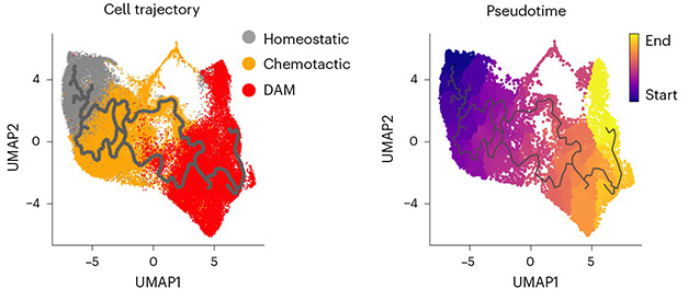
Do the VCAM1-positive microglia clear plaques, or just surround them and coax other phagocytotic cells to do the dirty work? Some microglia expressed both VCAM1 and DAM markers, hinting they were transitioning between the two states. Indeed, pseudotime analysis, which orders cells on a virtual timeline by their similarity in gene expression, suggested homeostatic microglia morph into chemotactic cells and then into DAMs (image below).

Morphing Microglia. Both cell trajectory (left) and pseudotime (right) analyses carved the same path for microglia in APP/PS1 mice. They move from homeostatic to chemotactic to DAM states. [Courtesy of Lau et al., Nature Aging, 2023.]
What within amyloid plaques was beckoning microglia? While Aβ is the primary component, plaques also contain lipids and other proteins, notably ApoE (Xiong et al., 2019; Dec 2021 news; Aug 2023 news). When Lau and Wu searched the STRING database of protein-protein interactions, they found that VCAM1 interacts with plaque-associated proteins, including ApoE, the CD44 receptor, and the integrin ITGB2.
To determine if any of the three interact with VCAM1 in vivo, the scientists injected microbeads coated with each protein into the mouse brains, injected IL-33, then analyzed microglial movement toward the beads 24 hours later. Only the ApoE-coated beads attracted the VCAM1-positive microglia. On the other hand, blocking ApoE with an antibody, or knocking it out genetically, not only prevented microglial migration toward plaques, but also their transition to DAMs and their ability to clear amyloid (image below). The results suggest that plaque-bound ApoE lures microglia and drives them to transition into a phagocytotic state.

No ApoE, No Migration. In the brains of APP/PS1 mice (left), phagocytic microglia (orange or arrowheads) congregate around plaques (blue). If an anti-ApoE antibody is injected into brain, the microglia (green) don’t budge (right). [Courtesy of Lau et al., Nature Aging, 2023.]
Are VCAM1-positive microglia important in AD? Postmortem analysis of brain tissue from 35 people who had had AD showed that VCAM1-expressing microglia surrounded amyloid plaques. In some cases, however, the number of microglia was small, suggesting an impaired response to amyloid. Why? Lau and Wu suspected a soluble version of VCAM1 (sVCAM1) may act as a decoy, competing with VCAM1-expressing cells for target receptors. Indeed, the people with few VCAM1-positive microglia surrounding plaques had more sVCAM1 in their cerebrospinal fluid. Scientists don’t know if sVCAM1 arises from cleavage of the membrane-bound form, or if it is made through alternative splicing.
Compared to healthy controls, people with AD had 34 percent more sVCAM1 in their blood, suggesting an uptick in VCAM1 processing. This mirrored high plasma levels of a soluble fragment of the IL-33 receptor, ST2. Ip previously reported that there is more soluble ST2 in AD plasma than in control blood (Jul 2022 news). “Interestingly, the two key regulatory axes of microglial chemotaxis—IL-33–ST2 and VCAM1 signaling—are dysregulated in AD,” the authors concluded.—Chelsea Weidman Burke
References
Research Models Citations
News Citations
- Hot DAM: Specific Microglia Engulf Plaques
- Amyloids Fibrillize, or Stay Solo, Based on Liaisons with Like Proteins
- Organization of Aβ Plaques and Tau Tangles Illuminated in AD Brain
- Receptor Decoy Raises Risk of Alzheimer’s—But Only in Women
Paper Citations
- Lau SF, Chen C, Fu WY, Qu JY, Cheung TH, Fu AK, Ip NY. IL-33-PU.1 Transcriptome Reprogramming Drives Functional State Transition and Clearance Activity of Microglia in Alzheimer's Disease. Cell Rep. 2020 Apr 21;31(3):107530. PubMed.
- Xiong F, Ge W, Ma C. Quantitative proteomics reveals distinct composition of amyloid plaques in Alzheimer's disease. Alzheimers Dement. 2019 Mar;15(3):429-440. Epub 2019 Jan 2 PubMed.
External Citations
Further Reading
No Available Further Reading
Primary Papers
- Lau SF, Wu W, Wong HY, Ouyang L, Qiao Y, Xu J, Lau JH, Wong C, Jiang Y, Holtzman DM, Fu AK, Ip NY. The VCAM1-ApoE pathway directs microglial chemotaxis and alleviates Alzheimer's disease pathology. Nat Aging. 2023 Oct;3(10):1219-1236. Epub 2023 Sep 21 PubMed.
Annotate
To make an annotation you must Login or Register.

Comments
Mayo Clinic
In this paper, Dr. Ip and colleagues extended their prior research on the role of interleukin-33 (IL-33) in microglial Aβ clearance. Their previous work had suggested that after injection in APP/PS1 mice IL-33 facilitates microglial Aβ clearance by initially inducing Aβ chemotaxis followed by Aβ phagocytosis (Lau et al., 2020). However, the precise pathway regulating this microglial response was unclear.
In this study, the researchers conducted a series of experiments to uncover these mechanisms. Through both bulk and single-cell transcriptomic analyses, they validated that, at the transcriptomic level, microglia transition from a homeostatic state to a chemotactic state, and subsequently to an Aβ-phagocytic state following IL-33 injection, consistent with their earlier findings. Furthermore, they identified a crucial role for VCAM1 in enabling microglia to acquire these chemotactic states after IL-33 injection. This was confirmed through experiments involving microglia-specific VCAM1 conditional knockout mice, which displayed an inability to respond to Aβ after IL-33 injection, among other supporting evidence. They also demonstrated that the injection of recombinant ApoE, not lipidated ApoE, could attract VCAM1+ microglia following IL-33 injection, and that the administration of an ApoE neutralizing antibody disrupted the IL-33-mediated migration of VCAM1+ microglia to Aβ plaques. These findings suggest that VCAM1+ microglia migrate toward Aβ plaques by sensing non-lipidated ApoE within these plaques. Finally, in humans, the team observed elevated soluble VCAM1 levels in both plasma and CSF among AD patients compared to control subjects. Additionally, CSF soluble VCAM1 levels showed a negative correlation with the presence of microglia surrounding Aβ plaques.
This paper presents a compelling connection between ApoE and VCAM1 in the IL-33-mediated microglial response to Aβ plaques. It also opens the door to several intriguing questions that merit further investigation. I think one critical area for exploration is the necessity for additional evidence elucidating how non-lipidated ApoE binds to VCAM1 and why lipidation of ApoE disrupts this interaction. It is also worth considering whether ApoE aggregation plays a role in this interaction, given that ApoE co-deposited with Aβ plaques is typically insoluble. Clarifying this distinction is essential for defining ApoE as a ligand for VCAM1. Another question is whether the ApoE-VCAM1 axis regulates microglial responses exclusively following IL-33 injection or if it represents a general mechanism by which microglia sense and respond to Aβ plaques. Drawing from Kim et al.'s work , it is worth noting that treatment with the ApoE neutralizing antibody (HJ6.3) appeared to significantly enhance microglial activity and reduce Aβ plaque load, contrasting the observations in this paper, where the administration of the same ApoE antibody reduced the microglial response to Aβ and abolished the IL-33-induced Aβ clearance (Kim et al., 2012). This suggests that the presence of IL-33 might alter ApoE-related microglial functions.
Overall, this outstanding paper by Dr. Ip and colleagues, along with the further questions it prompts, has the potential to significantly advance our understanding of ApoE and microglial biology in response to amyloid pathology.
References:
Lau SF, Chen C, Fu WY, Qu JY, Cheung TH, Fu AK, Ip NY. IL-33-PU.1 Transcriptome Reprogramming Drives Functional State Transition and Clearance Activity of Microglia in Alzheimer's Disease. Cell Rep. 2020 Apr 21;31(3):107530. PubMed.
Kim J, Eltorai AE, Jiang H, Liao F, Verghese PB, Stewart FR, Basak JM, Holtzman DM. Anti-apoE immunotherapy inhibits amyloid accumulation in a transgenic mouse model of Aβ amyloidosis. J Exp Med. 2012 Nov 19;209(12):2149-56. PubMed.
University of California San Diego
This looks like an interesting new role for VCAM1 in facilitating microglia migration to and phagocytosis of amyloid plaques. Although VCAM1 is well known to be involved in the interactions of monocytes and other immune cells with endothelial cells, this proposed function in microglia would be qualitatively different. The connection to ApoE is also interesting, but at present it is not clear whether, and if so how, VCAM1 directly detects ApoE. It will be of substantial interest to explore this aspect of the findings further and determine whether there are differences with respect to APOE3 and APOE4 alleles.
Mayo Clinic Florida
The APOE-TREM2 interaction is well established but APOE-VCAM1 may be an important co-stimulatory pathway for microglial recruitment to amyloid plaques. Indeed, AD patients with TREM2 R47H mutations still show recruitment of microglial to the amyloid plaque region and their hyperactivation (Korvatska et al., 2015; Sayed et al., 2021), although transplanted human TREM2 R47H iPSC-derived microglia show reduced plaque reactivity in the 5xFAD mouse model (Claes et al., 2021). These conflicting results may be due to the difference in the induction of the alternative pathway for the recruitment of microglia to the plaque region. Targeting VCAM1 may be an attractive alternative approach because treatment of AD cases with anti-TREM2 antibody was recently reported to induce ARIA (Aug 2023 news /news/conference-coverage/aria-inflammatory-reaction-vascular-amyloid).
References:
Korvatska O, Leverenz JB, Jayadev S, McMillan P, Kurtz I, Guo X, Rumbaugh M, Matsushita M, Girirajan S, Dorschner MO, Kiianitsa K, Yu CE, Brkanac Z, Garden GA, Raskind WH, Bird TD. R47H Variant of TREM2 Associated With Alzheimer Disease in a Large Late-Onset Family: Clinical, Genetic, and Neuropathological Study. JAMA Neurol. 2015 Aug;72(8):920-7. PubMed.
Sayed FA, Kodama L, Fan L, Carling GK, Udeochu JC, Le D, Li Q, Zhou L, Wong MY, Horowitz R, Ye P, Mathys H, Wang M, Niu X, Mazutis L, Jiang X, Wang X, Gao F, Brendel M, Telpoukhovskaia M, Tracy TE, Frost G, Zhou Y, Li Y, Qiu Y, Cheng Z, Yu G, Hardy J, Coppola G, Wang F, DeTure MA, Zhang B, Xie L, Trajnowski JQ, Lee VM, Gong S, Sinha SC, Dickson DW, Luo W, Gan L. AD-linked R47H-TREM2 mutation induces disease-enhancing microglial states via AKT hyperactivation. Sci Transl Med. 2021 Dec;13(622):eabe3947. PubMed.
Claes C, Danhash EP, Hasselmann J, Chadarevian JP, Shabestari SK, England WE, Lim TE, Hidalgo JL, Spitale RC, Davtyan H, Blurton-Jones M. Plaque-associated human microglia accumulate lipid droplets in a chimeric model of Alzheimer's disease. Mol Neurodegener. 2021 Jul 23;16(1):50. PubMed.
Make a Comment
To make a comment you must login or register.