Neuroprotective Extracellular Matrix Seen in C9ORF72 ALS/FTD Models
Quick Links
Amyotrophic lateral sclerosis and frontotemporal dementia are heterogeneous diseases that are difficult to fully model in mice. One of the more common forms is caused by hexanucleotide repeat expansion in the gene C9ORF72, which prompts both loss of function of the normal protein and expression of toxic polydipeptides. Since this discovery in 2011, scientists have modeled it in rodents nearly 20 different ways, by knocking out the gene, or by expressing various combinations of polydipeptide repeats. None model all aspects of ALS or FTD; for a review see Todd and Petrucelli, 2022.
- Scientists created two new models of C9ORF72 ALS/FTD.
- The mice express half of the normal C9ORF72 and polydipeptide repeats.
- They reflect certain aspects of the disease, such as motor neuron degeneration.
- Their spinal cords overproduce collagens, likely to protect neurons.
Now, scientists led by Elizabeth Fisher and Adrian Isaacs of University College London have created two new knock-in models combining C9ORF72 loss-of-function and specific dipeptide expression. In the February 29 Nature Neuroscience, they reported that these mice capture some aspects of ALS/FTD, such as hyperactive motor cortex neurons, spinal motor neuron loss with age, and worsening coordination. Unexpectedly, their spinal cords—driven by TGF-β1—cranked up production of extracellular matrix proteins.
“This represents another advance in C9ORF72 modeling,” wrote Robert Baloh of Novartis. “None of the existing rodent models appear to be significantly better than the others, but each may represent a piece of the puzzle, and the broad selection of different models now available allows groups to test different therapeutic approaches in an in vivo setting.”
First author Carmelo Milioto and colleagues generated mice expressing largely physiological levels of one copy of wild-type murine C9ORF72 and another copy with a stretch of DNA encoding 400 repeats of poly-glycine-arginine (polyGR) or poly-proline-arginine (polyPR) inserted right after the C9ORF72 start codon. More than 30 repeats are pathogenic, and most people with ALS/FTD have hundreds (Sep 2011 news). PolyGR and polyPR are two of five repeat types that form neuronal inclusions in ALS/FTD, and these two seem most toxic in human cells, mice, and fruit flies (Feb 2013 news; Dec 2014 news; Jun 2018 news).
By 3 months old, (GR)400 and (PR)400 mice expressed 40 percent less C9ORF72 protein in their brains and spinal cords. They also made polyGR or polyPR, the former at levels similar to that in cortical tissue from ALS/FTD patients. (The scientists did not compare polyPR to human tissue.) However, only polyGR diffusely stained neurons in mouse cortical and spinal cord slices. Isaacs believes both polydipeptides were soluble, rather than forming inclusions, and the polyGR antibody was able to detect the soluble form while the polyPR one did not.
Did the polydipeptides affect neuronal function? In vivo calcium imaging revealed more neuronal and network hyperexcitability in the motor cortices of 18-month-old (GR)400 mice than in wild-types. (PR)400 mice had no more overactive neurons than wild-type, suggesting a biological difference between the two peptides, Isaacs said. Neither model had cortical neurodegeneration.
However, in the lumbar spine, neurons withered. While 6-month-old mice had as many lower motor neurons as wild-types, by 12 months they had 20 percent fewer. Up to 40 percent of motoneurons innervating the leg muscles did not fire when stimulated with an electrode. This translated to declining motor function starting at 6 months, with mice falling off a rotarod more quickly than wild-types.
Even so, these mice did not recapitulate all aspects of ALS/FTD. Neither had any astro- or microgliosis or TDP-43 mislocalization to the neuronal cytoplasm.

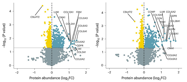
Copious Collagen. In spinal cord tissue from (GR)400 (left) and (PR)400 (right) mice, C9ORF72 was downregulated (yellow). Extracellular matrix proteins, including many collagens, were upregulated (blue). [Courtesy of Milioto et al., Nature Neuroscience, 2024.]
What was happening at a cellular level? Milioto et al. analyzed proteomics in bulk tissue homogenates from 12-month-old (GR)400 and (PR)400 mice. Both expressed fewer synaptic proteins in the spinal cord but not cortex, mirroring the neuron loss. The mice upregulated extracellular matrix (ECM) proteins and the ECM upstream regulator TGF-β1 (image above). Spinal cord, but not cortical, neurons contained 50 percent more of the type VI collagen component COL6A1, one of the most upregulated ECM proteins.
Isaacs had not seen this ECM signature in ALS/FTD models before. When perusing previously published proteomic data from motor neurons of people with ALS, he noticed that they, too, had upregulated ECM proteins and TGF-β1 (NeuroLINCS Consortium, 2021; Highley et al., 2014). Given that ECM proteins were only upregulated in areas where (GR)400 or (PR)400 mice had neurodegeneration, Isaacs thought the ECM response might try to protect neurons.
To find out, the authors turned to fruit flies with 36 polyGR repeats. They develop eye degeneration, a convenient readout of neurodegeneration. Indeed, the flies upregulated orthologues of COL6A1 and TGF-β1 genes. Knocking down either worsened eye degeneration, and the flies died sooner (image below).

No More Eye. Compared to eyes of wild-type fruit flies (left), those of flies with polyGR repeats degenerate (second panel). They disappear if orthologues of the TGFB1 (third panel) or COL6A1 (right) genes are knocked down. [Courtesy of Milioto et al., Nature Neuroscience, 2024.]
Mice expressing human amyloid precursor protein also overproduce TGF-β1 and COL6A1, which protect their neurons from Aβ toxicity (Jan 2009 news). This hints that the ECM pathway might be broadly neuroprotective. “It's an open question whether activating it, either via TGF-β1 or COL6A1, would provide protection in people,” Isaacs told Alzforum.
The (GR)400 and (PR)400 mice will be freely available to researchers through the European Mutant Mouse Archive.—Chelsea Weidman Burke
References
News Citations
- Corrupt Code: DNA Repeats Are Common Cause for ALS and FTD
- Second Study Sees Intron in FTLD Gene Translated
- Live-Cell Studies Blame Arginine Peptides for C9ORF72’s Crimes
- In New ALS/FTD Mouse Model, Poly(GR) Peptides Poison Ribosomes
- Sticking It to Oligomers—Does Collagen Protect Neurons From Aβ?
Paper Citations
- Todd TW, Petrucelli L. Modelling amyotrophic lateral sclerosis in rodents. Nat Rev Neurosci. 2022 Apr;23(4):231-251. Epub 2022 Mar 8 PubMed.
- NeuroLINCS Consortium, Li J, Lim RG, Kaye JA, Dardov V, Coyne AN, Wu J, Milani P, Cheng A, Thompson TG, Ornelas L, Frank A, Adam M, Banuelos MG, Casale M, Cox V, Escalante-Chong R, Daigle JG, Gomez E, Hayes L, Holewenski R, Lei S, Lenail A, Lima L, Mandefro B, Matlock A, Panther L, Patel-Murray NL, Pham J, Ramamoorthy D, Sachs K, Shelley B, Stocksdale J, Trost H, Wilhelm M, Venkatraman V, Wassie BT, Wyman S, Yang S, NYGC ALS Consortium, Van Eyk JE, Lloyd TE, Finkbeiner S, Fraenkel E, Rothstein JD, Sareen D, Svendsen CN, Thompson LM. An integrated multi-omic analysis of iPSC-derived motor neurons from C9ORF72 ALS patients. iScience. 2021 Nov 19;24(11):103221. Epub 2021 Oct 12 PubMed.
- Highley JR, Kirby J, Jansweijer JA, Webb PS, Hewamadduma CA, Heath PR, Higginbottom A, Raman R, Ferraiuolo L, Cooper-Knock J, McDermott CJ, Wharton SB, Shaw PJ, Ince PG. Loss of nuclear TDP-43 in amyotrophic lateral sclerosis (ALS) causes altered expression of splicing machinery and widespread dysregulation of RNA splicing in motor neurones. Neuropathol Appl Neurobiol. 2014 Oct;40(6):670-85. PubMed.
External Citations
Further Reading
Papers
- Meroni M, Crippa V, Cristofani R, Rusmini P, Cicardi ME, Messi E, Piccolella M, Tedesco B, Ferrari V, Sorarù G, Pennuto M, Poletti A, Galbiati M. Transforming growth factor beta 1 signaling is altered in the spinal cord and muscle of amyotrophic lateral sclerosis mice and patients. Neurobiol Aging. 2019 Oct;82:48-59. Epub 2019 Jul 10 PubMed.
Primary Papers
- Milioto C, Carcolé M, Giblin A, Coneys R, Attrebi O, Ahmed M, Harris SS, Lee BI, Yang M, Ellingford RA, Nirujogi RS, Biggs D, Salomonsson S, Zanovello M, de Oliveira P, Katona E, Glaria I, Mikheenko A, Geary B, Udine E, Vaizoglu D, Anoar S, Jotangiya K, Crowley G, Smeeth DM, Adams ML, Niccoli T, Rademakers R, van Blitterswijk M, Devoy A, Hong S, Partridge L, Coyne AN, Fratta P, Alessi DR, Davies B, Busche MA, Greensmith L, Fisher EM, Isaacs AM. PolyGR and polyPR knock-in mice reveal a conserved neuroprotective extracellular matrix signature in C9orf72 ALS/FTD neurons. Nat Neurosci. 2024 Apr;27(4):643-655. Epub 2024 Feb 29 PubMed.
Annotate
To make an annotation you must Login or Register.

Comments
Director of Neuromuscular Medicine, Cedars-Sinai Medical Center
This represents another advance in C9ORF72 modeling, by placing expression of one of two different artificially engineered DPR proteins (GR or PR) under the control of the endogenous C9ORF72 promoter. Interestingly, the authors found that lower motor neurons were particularly vulnerable in these mice, while in humans, DPR proteins are only rarely detected in spinal motor neurons and their relevance in disease has therefore been questioned (Gomez-Deza et al., 2015).
The jury is still out on whether rodent models will be helpful in predicting which DPRs are pathogenic in human ALS/FTD, as others have also observed differences such as polyGA appearing to be more toxic than polyPR, despite the fact that the PR is typically more toxic in vitro (LaClair et al., 2020).
It is safe to say that none of the existing rodent models appears to be significantly better than the others. Each may represent a piece of the puzzle, and the broad selection of different models now available allows groups to test different therapeutic approaches in an in vivo setting.
References:
Gomez-Deza J, Lee YB, Troakes C, Nolan M, Al-Sarraj S, Gallo JM, Shaw CE. Dipeptide repeat protein inclusions are rare in the spinal cord and almost absent from motor neurons in C9ORF72 mutant amyotrophic lateral sclerosis and are unlikely to cause their degeneration. Acta Neuropathol Commun. 2015 Jun 25;3:38. PubMed.
LaClair KD, Zhou Q, Michaelsen M, Wefers B, Brill MS, Janjic A, Rathkolb B, Farny D, Cygan M, de Angelis MH, Wurst W, Neumann M, Enard W, Misgeld T, Arzberger T, Edbauer D. Congenic expression of poly-GA but not poly-PR in mice triggers selective neuron loss and interferon responses found in C9orf72 ALS. Acta Neuropathol. 2020 Aug;140(2):121-142. Epub 2020 Jun 19 PubMed.
Amsterdam UMC, VU University
Vrije Universiteit Amsterdam
VU University Medical Center
We have read the study from Milioto et al. with much interest, because last year we published a clinical study in which we made a similar observation (Gogishvili et al., 2023), namely that while TGF-β1 was increased in dementia compared to controls, it was lower (at normal levels) in the dementia patients with fast cognitive decline.
The aim of our study was to identify CSF proteins biomarkers that predict fast cognitive decline within dementia patients. TGF-β1 was one of the identified markers with the best predictive power. Therefore, we hypothesized that increased levels of TGF-β1 in CSF in dementia might be a protective response to the pathology. The current study supports this hypothesis and demonstrates how this mechanism could work in their C9ORF72 model. Very exciting!
It is also an illustration of the potential of combining results from CSF and experimental studies to better understand the disease mechanisms, and how that could help us to ultimately capture these processes in humans.
References:
Gogishvili D, Vromen EM, Koppes-den Hertog S, Lemstra AW, Pijnenburg YA, Visser PJ, Tijms BM, Del Campo M, Abeln S, Teunissen CE, Vermunt L. Discovery of novel CSF biomarkers to predict progression in dementia using machine learning. Sci Rep. 2023 Apr 21;13(1):6531. PubMed.
Make a Comment
To make a comment you must login or register.