Paper Alert: Centiloid Scale Aims to Unify Amyloid PET
Quick Links
Several ligands, with different fibrillar Aβ-binding characteristics, are being used to image amyloid accumulation in the human brain. To help researchers compare data across studies and among labs, scientists led by William Klunk at the University of Pittsburgh began the Centiloid Project, an effort to standardize amyloid imaging measurements from positron emission tomography (PET) scans. In the October 28 Alzheimer’s and Dementia, the scientists laid out their method for fitting results to a common scale, regardless of tracer, brain reference region, or method of analysis used. With centiloids, scientists will be able to translate their own data to a common standard and directly compare amyloid-imaging measurements. “The ultimate goal is to have everybody speaking the same language, while leaving them free to carry out their own protocols,” Klunk told Alzforum. He presented on this initiative at the 2013 Human Amyloid Imaging conference (see Feb 2013 news story).

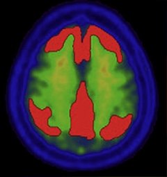
The standard volume of interest for PiB scans (red), which is used to construct the centiloid scale. [Reprinted from Klunk et al., with permission from Elsevier.]
With this publication, Klunk and colleagues have completed the first of three steps toward a common imaging standard. They used Pittsburgh Compound B (PiB) PET scans of 34 young, healthy controls and 47 AD patients to define the bottom and top of the centiloid scale, respectively. Taking PET scans 50 to 70 minutes after PiB injection, the researchers measured Aβ in a cortical region that accumulates the most amyloid, using the whole cerebellum as a reference (see image at left). Data from the youngest volunteers, who should have accumulated no Aβ, determined the zero point on the scale, while the most advanced AD patients, who were assumed to have brains saturated with the peptide, served as the 100-point reference. Klunk and colleagues then assigned each individual scan a global centiloid value based on the new scale. The centiloid scale serves as a standard to which future amyloid imaging data can be converted, enabling scientists to directly compare results among studies.
In order to express their own PiB data in centiloids, researchers must first confirm that their methods of analysis generate the same standard uptake value ratios (SUVrs) as did Klunk and colleagues’. For this purpose, the original raw scans Klunk used to generate the centiloid scale are available through the Global Alzheimer's Association Interactive Network (GAAIN). SUVrs within 2 percent of the originals demonstrate that the same centiloids can be derived.
If scientists want to use a different tracer, they’ll need to convert their data to equivalent PiB units, which can then be used to obtain centiloid values. Klunk and colleagues have not done this themselves. They expect other researchers will step in. The ligand of choice will have to be directly compared to PiB by scanning 25 people with both tracers within a three-month period. Klunk recommends imaging 10 healthy controls, five AD patients, and 10 people likely to have intermediate levels of amyloid. The head-to-head comparison will allow researchers to convert ligand SUVrs to equivalent PiB SUVrs, and then to centiloids. This calibration step would only have to be completed once per tracer, then other groups could use this same calibration curve.
In some cases, researchers may want to use another methodology for a tracer that has been calibrated already—for example, employing a different reference region or time from injection. In that case, they will not need to acquire new scans, but can simply calibrate their method using the available raw data.
Klunk and colleagues ask that any group that completes calibration steps for a given tracer, or method, make the data publicly available. That will enable step three, in which scientists simply download published calibration data that matches their own methods, demonstrate that they can replicate the calculations with their analytical tools, and express their own data in centiloids.
How widely will this process be used? Klunk predicted that manufacturers of F18-labeled tracers could calibrate these ligands in relatively short order. After that, multicenter studies wishing to compare data across centers and tracers could use the scale. Eventually, researchers may publish both in their own units and in centiloids, to make data comparable among studies, Klunk said. He has already received emails from researchers wondering how to convert their measurements. In terms of comparing new F18-labeled tracers to PiB, no published data are available yet, though co-author Robert Koeppe, University of Michigan, Ann Arbor, said that those comparisons are underway.
“Overall the project is a highly welcome initiative,” Zsolt Cselényi, of AstraZeneca, at the Karolinska Institutet, Stockholm, wrote to Alzforum in an email (see full comment below). “The ability to confidently compare and translate quantitative results among the various amyloid tracers should facilitate both basic research on disease pathophysiology and the development of drugs acting on the amyloid pathway.”—Gwyneth Dickey Zakaib
References
News Citations
External Citations
Further Reading
Papers
- Landau SM, Thomas BA, Thurfjell L, Schmidt M, Margolin R, Mintun M, Pontecorvo M, Baker SL, Jagust WJ, Alzheimer’s Disease Neuroimaging Initiative. Amyloid PET imaging in Alzheimer's disease: a comparison of three radiotracers. Eur J Nucl Med Mol Imaging. 2014 Jul;41(7):1398-407. Epub 2014 Mar 20 PubMed.
Primary Papers
- Klunk WE, Koeppe RA, Price JC, Benzinger TL, Devous MD Sr, Jagust WJ, Johnson KA, Mathis CA, Minhas D, Pontecorvo MJ, Rowe CC, Skovronsky DM, Mintun MA. The Centiloid Project: standardizing quantitative amyloid plaque estimation by PET. Alzheimers Dement. 2015 Jan;11(1):1-15.e1-4. Epub 2014 Oct 28 PubMed.
Annotate
To make an annotation you must Login or Register.

Comments
AstraZeneca
Overall, this project is a highly welcome initiative. The ability to confidently compare and translate quantitative results among the various amyloid tracers should facilitate both basic research on disease pathophysiology and the development of drugs acting on the amyloid pathway. Large, multicenter research or trials could confidently employ different amyloid ligands depending on availability, approval status, or other factors, without having to worry about the difficulties of pooling results together in the end. In theory, even longitudinal follow-up of a given patient could make use of different ligands at different time points and still arrive at a numerical value quantifying changes in amyloid load. A welcome “side-effect” of the centiloid scale is that measures of reliability will be more comparable between tracers, which is very important when calculating power or sample sizes for trials planning to use a mixed set of ligands.
However, there are some challenges that the project needs to overcome. The first is related to the inherent convertibility of amyloid measures between the ligands. The centiloid scale is essentially a linear scale anchored at two points. Convertibility requires, among other things, that the amyloid load estimates for all ligands should change in a similar, linear way in response to increasing true plaque load. That is to say, non-linearities in the dynamic range may lead to problems of converting/standardizing intermediate load levels. Some possible sources of non-linearity are:
These issues may be particularly important in case of longitudinal tracking of amyloid load and less so, for example, when simply separating amyloid positives and negatives. A further challenge can be to try to apply centiloid standardization at the regional/voxel level. While the project seems geared toward standardization of the global load measure, it may be interesting to use it also on the region/image level, and in that case the above-described and similar non-linearities (especially differences in white-matter binding) may make it difficult to obtain an equivalent centiloid image.
Another set of challenges come at the time of trying to implement/use the centiloid scale in actual research projects/drug trials:
- On-site calibration is key to ensure comparability of published findings. This step is described in the Centiloid Project. It is worth emphasizing that correct implementation will be facilitated by detailed description of acquisition, reconstruction settings/processing methodologies used to determine the values for the public calibration data sets (such as image preprocessing steps with important parameters, e.g., smoothing filters used, derivation of regions of interest, quantification method, etc.).
- It may be envisaged that research groups/pharmaceutical companies would simply adopt using standard acquisition protocols and methods (cookbook recipes) instead of full local (re-) calibration work. This puts even stronger demands on having fully documented descriptions of settings/methods used for the calibration data sets. Ideally, even the actual software components or algorithms could be shared so that groups might simply and reliably analyze their data without on-site calibration.
View all comments by Zsolt CselényiMake a Comment
To make a comment you must login or register.