Spooned by a Fold: MK-6240 Nestles Within Tau Protofilament
Quick Links
In the decade since the first tau PET ligands burst onto the scene, chemists have been tinkering with the compounds to enhance their penchant for pathological tau. A study posted September 22 on bioRxiv reveals just how snugly one of the resulting second-generation tracers—MK-6240—fits its target. Scientists led by Sarah Shahmoradian and Marc Diamond of University of Texas Southwestern Medical Center in Dallas and Pedro Rosa-Neto of McGill University in Montreal wielded cryo-EM to visualize how tau fibrils embraced the ligand. For every tau monomer wound into the fibril, a single MK-6240 molecule nestled cozily within the cavity of the C-shaped protofilament core. What’s more, the tracer molecules aligned with one another within the fibril, such that they interacted with each other even more than they did with their target. The findings hint at an explanation behind this tracer’s high avidity, and specificity, for tau fibrils that form in AD.
- Cryo-EM resolved structure of MK-6240 bound to tau.
- The ligand bound to the inner cavity of each C-shaped protofilament.
- Stacked MK-6240 molecules interact more with each other than with the tau filament.
First author Peter Kunach and colleagues extracted tau filaments from the frontal cortex of a person who had died with AD, and then incubated the fibrils with a high concentration of the tracer. Cryo-EM revealed characteristic AD paired helical filaments (PHFs), with their stacks of back-to-back C-shaped protofilaments. In samples incubated with the tracer, each C clutched an additional density within a tiny inlet bordered by residues Q351 and I360 (first image below). At a 1:1 stoichiometry with tau monomers, MK-6240 molecules were stacked at 4.8Å apart in a slanted, staggered arrangement along the fibril column.

Perfect Fit. A cross-sectional view of a paired helical filament of tau reveals the MK-6240 tracer (red, top; or brown, bottom) held snugly within the cavity of each C-shaped protofilament. [Courtesy of Kunach et al., BioRxiv, 2023]
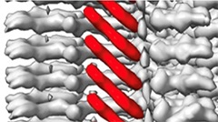
Another striking feature was the close alignment of the tracer molecules within the fibril. Stacked diagonally, and proximal to each other down the fibril axis, the ligands appeared to form a molecular glue (see image below). In fact, the surface area involved in these ligand-ligand interactions was greater than that between the ligand and its target protein. Notably, Genentech’s GTP-1 tau tracer was recently reported to bind in a similar manner (Merz et al., 2023). This arrangement could explain why these tracers bind with such high avidity to tau PHFs, the authors proposed.
“These studies help people understand the special binding mode for GTP-1 and MK-6240 in AD tau PHFs, and provide structural basis for further computational based tracer design,” commented Junhao Li and Hans Agren of Uppsala University in Sweden.

PET Tracer Glue? Individual molecules of MK-6240 stack close and diagonally along the helical axis of AD tau filaments. [Courtesy of Kunach et al., BioRxiv, 2023.]
Tau structure aficionados Sjors Scheres and Michel Goedert of the MRC Laboratory of Molecular Biology at Cambridge, U.K., were unable to visualize the first-generation tracer, flortaucipir, buddied up with AD PHFs, but curiously, they did spy it bound to tau filaments twisted into a chronic traumatic encephalopathy (CTE) fold (Shi et al., 2023). Flortaucipir bound to these CTE tau fibrils within the more open curve of their C-shaped protofilaments.
In a joint comment to Alzforum, Scheres and Goedert called the new high-resolution structure of MK-6240 bound to AD tau PHFs beautiful. However, they noted, as did the authors, that it remains to be seen if the same binding arrangement occurs during PET imaging, when tracer concentrations are much lower (comment below). Chet Mathis of the University of Pittsburg in Pennsylvania raised the same question, noting that tracer concentrations in the brain would be 20,000-fold lower than the conditions used in related studies. At these lower concentrations, the ligand-ligand interactions, aka “molecular glue,” would be statistically unlikely to form, he wrote. “It is possible that the high affinity PET radioligand binding site for [18F]MK-6240 on tau PHFs/neurofibrillary tangles is in the C-shaped cavity containing amino acids Q351-I360, but it is also possible that it resides in a location where ligand-ligand interactions are minimal, do not dominate, and would therefore not be detectable using cryo-EM,” he wrote.
Shahmoradian told Alzforum that they are investigating the binding mode of the tracer at sub-stoichiometric conditions, where tau monomers outnumber tracer molecules. Their preliminary data confirm the existence of stacked binding modes that occur at higher tracer concentrations, she said.—Jessica Shugart
References
Paper Citations
- Merz GE, Chalkley MJ, Tan SK, Tse E, Lee J, Prusiner SB, Paras NA, DeGrado WF, Southworth DR. Stacked binding of a PET ligand to Alzheimer's tau paired helical filaments. Nat Commun. 2023 May 26;14(1):3048. PubMed.
- Shi Y, Ghetti B, Goedert M, Scheres SH. Cryo-EM Structures of Chronic Traumatic Encephalopathy Tau Filaments with PET Ligand Flortaucipir. J Mol Biol. 2023 Jun 1;435(11):168025. Epub 2023 Jun 16 PubMed.
Further Reading
Papers
- Xia CF, Arteaga J, Chen G, Gangadharmath U, Gomez LF, Kasi D, Lam C, Liang Q, Liu C, Mocharla VP, Mu F, Sinha A, Su H, Szardenings AK, Walsh JC, Wang E, Yu C, Zhang W, Zhao T, Kolb HC. [(18)F]T807, a novel tau positron emission tomography imaging agent for Alzheimer's disease. Alzheimers Dement. 2013 Feb 11; PubMed.
Primary Papers
- Kunach P, Vaquer-Alicea J, Smith MS, Hopewell R, Monistrol J, Moquin L, Therriault J, Tissot C, Rahmouni N, Massarweh G, Soucy J-P, Guiot M-C, Shoichet BK, Rosa-Neto PK, Diamond MI, Shahmoradian SH. Cryo-EM structure of Alzheimer disease tau filaments with PET ligand MK-6240. 2023 Sep 22 10.1101/2023.09.22.558671 (version 1) bioRxiv.
Annotate
To make an annotation you must Login or Register.

Comments
MRC Laboratory of Molecular Biology
MRC Laboratory of Molecular Biology
This preprint by Kunach et al. describes a beautiful 2.1 Å resolution cryo-EM structure of PHFs from the frontal cortex of an individual with sporadic Alzheimer’s disease in complex with the PET ligand MK-6240. It represents the fourth cryo-EM structure of tau filaments in complex with a PET ligand. The compounds APN-1607 (Shi et al., 2021), flortaucipir (Shi et al., 2023), and GTP-1 (Merz et al., 2023) have been visualised previously. The binding mode of MK-6240, which stacks with full occupancy relative to the tau monomers, and with a tilted orientation with respect to the helical axis to allow optimal stacking of the aromatic compound, resembles that of GTP-1 binding to tau PHFs and of flortaucipir binding to CTE type I filaments. As also discussed in this paper, it remains unclear whether this binding mode is the same as the binding mode that is relevant to imaging in vivo. In this context, it is of note that similar experiments with flortaucipir, which is an FDA-approved PET ligand for tau inclusions of Alzheimer’s disease, did not reveal binding to PHFs. It is possible that the high concentration of PET ligands used in these studies could result in the visualisation of binding modes that are not relevant in vivo. The authors propose to address this question by directing future work toward solving structures using sub-stoichiometric conditions.
References:
Shi Y, Murzin AG, Falcon B, Epstein A, Machin J, Tempest P, Newell KL, Vidal R, Garringer HJ, Sahara N, Higuchi M, Ghetti B, Jang MK, Scheres SH, Goedert M. Cryo-EM structures of tau filaments from Alzheimer's disease with PET ligand APN-1607. Acta Neuropathol. 2021 May;141(5):697-708. Epub 2021 Mar 16 PubMed. Correction.
Shi Y, Ghetti B, Goedert M, Scheres SH. Cryo-EM Structures of Chronic Traumatic Encephalopathy Tau Filaments with PET Ligand Flortaucipir. J Mol Biol. 2023 Jun 1;435(11):168025. Epub 2023 Jun 16 PubMed.
Merz GE, Chalkley MJ, Tan SK, Tse E, Lee J, Prusiner SB, Paras NA, DeGrado WF, Southworth DR. Stacked binding of a PET ligand to Alzheimer's tau paired helical filaments. Nat Commun. 2023 May 26;14(1):3048. PubMed.
PET Facility
Ligand-ligand interactions appear to dominate binding energetics at very high ligand concentrations, and are a consistent finding with cryo-EM studies of ligand-fibril interactions, such as those reported with MK-6240 and paired helical filaments (PHF) in this study. Still, it is important to note that significant radioligand-radioligand interactions at the high affinity PHF binding site are statistically unlikely at the concentrations employed in PET imaging studies, which are typically conducted at 20,000-fold lower ligand concentrations than utilized in this study i.e., <1 nM versus 20 μM used here. It is possible that the high-affinity, PET radioligand binding site for [18F]MK-6240 on PHF/neurofibrillary tangles is in the C-shaped cavity containing amino acids Q351-I360, but it is also possible that it resides in a location where ligand-ligand interactions are minimal, do not dominate, and would therefore not be detectable using cryo-EM.
Uppsala University
Together with a recent study that solved the structure of GTP1 in complex with tau PHFs from an AD brain sample (Merz et al., 2023), now we have the CryoEM structure for the AD tau PHF:MK6240 complex, which is also obtained with a 1:1 stoichiometry. We can see that the stacking of GTP1 and MK6240 both induce significant conformational change for the binding site, spanning from Q351 to I360. For example, the binding of GTP1 and MK6240 significantly enhanced the formation of the salt bridge between K353 and D358 compared to the apo AD tau PHFs (PDB 6HRE). These studies help people understand the special binding mode for GTP1 and MK6240 in AD tau PHFs and provide structural bases for further computational-based tracer design that can account for the aspects of dynamics, affinities, and kinetics.
Note that both GTP1 and MK6240 are not so linear in shape compared to PBB3/PMPBB3 (APN1607). In a recent paper, the stacked binding mode was also observed for the linear F0502B, containing a medium aliphatic side chain, in α-synuclein fibrils (Xiang et al., 2023). However, the concentration of F0502B was much higher than GTP1 and MK6240. An intuition from these studies is that the more linear the tracer, the more unlikely it is to adopt the self-stacked binding mode. This assumption may be supported by the CroEM density map of APN1607 in AD tau fibrils (Shi et al., 2021).
References:
Merz GE, Chalkley MJ, Tan SK, Tse E, Lee J, Prusiner SB, Paras NA, DeGrado WF, Southworth DR. Stacked binding of a PET ligand to Alzheimer's tau paired helical filaments. Nat Commun. 2023 May 26;14(1):3048. PubMed.
Xiang J, Tao Y, Xia Y, Luo S, Zhao Q, Li B, Zhang X, Sun Y, Xia W, Zhang M, Kang SS, Ahn EH, Liu X, Xie F, Guan Y, Yang JJ, Bu L, Wu S, Wang X, Cao X, Liu C, Zhang Z, Li D, Ye K. Development of an α-synuclein positron emission tomography tracer for imaging synucleinopathies. Cell. 2023 Aug 3;186(16):3350-3367.e19. Epub 2023 Jul 7 PubMed.
Shi Y, Murzin AG, Falcon B, Epstein A, Machin J, Tempest P, Newell KL, Vidal R, Garringer HJ, Sahara N, Higuchi M, Ghetti B, Jang MK, Scheres SH, Goedert M. Cryo-EM structures of tau filaments from Alzheimer's disease with PET ligand APN-1607. Acta Neuropathol. 2021 May;141(5):697-708. Epub 2021 Mar 16 PubMed. Correction.
Make a Comment
To make a comment you must login or register.