Transparent Bodies Allow Neural Networks to ‘Apparate’
Quick Links
It’s not Harry Potter magic. But all the same, in the July 31 Cell, researchers describe a technique that allows whole cellular networks to come into view in situ. The technique turns entire rodents see-through and permeable, granting open optical access to every nook, cranny, and neuron in the body. Called “whole-body clearing,” the method strips away the lipids and membranes that normally limit the depths to which light from microscopes can probe. With this technique, researchers can trace neural networks within the brain outward to those that extend to organs throughout the body, and avoid the painstaking procedure of imaging thin slices of tissue one at a time. The whole-body technique one-ups previous studies in which researchers made an entire rodent brain transparent.
“There is a very large effort to understand the brain connectome; however, there is very little known about the peripheral nervous system,” senior author Viviana Gradinaru of the California Institute of Technology in Pasadena told Alzforum. Understanding the diverse neural networks that control vital organs will provide a better foundation for learning how best to treat all manner of diseases, she added. For that to happen, researchers need the ability to image vast stretches of networks that run from the brain to organs and back, a task too daunting for researchers to perform slice by slice.

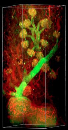
Unimpeded view.
Antibodies specific for tubulin (green) and DRAQ5 (red) label kidney glomeruli after being delivered systemically into a clear mouse through the PARS technique. [Image courtesy of Yang et al., Cell, 2014.]
The concept of tissue clearing, which involves destroying lipid membranes with detergents to render the tissue transparent, is nothing new. Alas, older techniques often damaged tissue or altered its morphology. More recent techniques have managed to keep tissues intact, but the chemicals used for clearing made the tissue impermeable to staining reagents such as fluorescently tagged antibodies. Limitations in the working depth of microscopes also stymied motivation to create organ- or body-clearing methods amenable to imaging, according to Gradinaru. “With recent advances in microscopy and imaging, I would say the time is prime for this technique now,” she said.
As a staff scientist at Stanford University, Gradinaru helped develop the CLARITY method, which made a rodent brain transparent and permeable enough to allow access to antibodies for staining (see Apr 2013 news story). In that study, researchers embedded the brain in a polymerizing gel, then used an electrical current to promote the distribution of detergents throughout the tissue, rendering it transparent. The problem with that method, Gradinaru said, was that if the conditions were not “just right,” the current could burn the tissue.
Wanting to create both a gentler clearing method and render an entire body transparent, first author Bin Yang and colleagues started off by tweaking their gel and detergent solutions and testing them on tissue slices. The researchers settled on a reagent combination that created a gel with pores large enough for detergent to pass through without the added electrical jolt. Using this “Goldilocks” protocol, which the researchers dubbed PACT (passive CLARITY), they stained and imaged 1- to 3-mm thick slices of brain, kidney, heart, lung, and intestine. Besides various tissue-specific antibodies and dyes, the researchers also labeled mRNA in cleared brain slices with fluorescent oligonucleotide probes. Uncleared brain slices had high background noise due to tissue auto-fluorescence; in contrast, the transparent slices enabled visualization of single molecules of β-actin mRNA. Gradinaru hopes the technique will one day facilitate the analysis of global gene expression throughout organs.
To scale up the PACT method to the entire rodent body, the researchers exploited the vasculature to deliver hydrogel and clearing solutions. Using a pump connected to a cardiac catheter, the researchers cycled clearing solutions throughout mice and rats for one or two weeks, respectively, until the rodents’ entire bodies became transparent. The researchers then delivered fluorescent antibodies and dyes via the same perfusion system to the whole body for imaging. The researchers called this method PARS (perfusion-assisted agent release in situ). Using it in mice expressing enhanced green fluorescent protein (eGFP) under the Thy1 promoter, the researchers were able to peer through standard confocal microscopes deep into the centers of multiple organs—including brain, spinal cord, kidney, lung, liver, and pancreas—and discern subcellular detail.
The blood-brain barrier remained a formidable foe. Despite clearing, it still prevented efficient delivery of full-size antibodies to the rodent brain, Gradinaru said. However, when the researchers used PARS to deliver GFAP-specific nanobodies—single-domain antibody fragments about one-tenth the size of full antibodies—glial cells in the brain vasculature and in surrounding tissue were extensively labeled. Unfortunately, nanobodies are not yet available for the full complement of brain proteins, Gradinaru said. Once they are, nanobodies will greatly expand the utility of the PARS technique to access the brain along with other organs in the body.
To get around this limitation now, the researchers developed a brain-specific PARS technique—PARS-CSF—in which they cycled clearing fluids directly into the cerebrospinal fluid in different areas of interest throughout the central nervous system. That way, the researchers cleared the brain or spinal cord in four days. PARS-CSF could be useful for researchers who want to label different regions of the brain or spinal cord without clearing the entire body, Gradinaru said.

Down to the Details. CA1 neurons in the hippocampus retain their delicate structure after PARS-CSF clearing in a mouse that had previously been infected with AAV9-eGFP. [Image courtesy of Yang et al., Cell 2014.]
Ashish Raj of Weill Cornell Medical College in New York called the study beautiful. “This could be invaluable for animal models of Alzheimer’s, for instance, where one wants to see how pathology and atrophy progress over time,” he wrote in an email to Alzforum (see full comment below).
Gradinaru plans to use the new techniques to trace degeneration of cells throughout the brain and body of rodent models of disease. Tracking which cells harbor pathogenic proteins or show signs of death throughout the course of a disease such as Parkinson’s, Gradinaru said, could prove useful in understanding how and where the disease originates and spreads. Some hypothesize that Parkinson’s disease originates in the gut and spreads to the brain via the vagus nerve, a complex nerve with myriad fibers (see Jul 2011 news series). The vagus nerve also has been implicated in depression, Gradinaru said, so taking a whole-body approach would be particularly useful in understanding how this complex nerve mediates disorders. She said the ultimate goal is to understand such networks well enough to target them with electrical stimulation in just the right spots to treat disease. —Jessica Shugart
References
News Citations
Further Reading
Papers
- Birmingham K, Gradinaru V, Anikeeva P, Grill WM, Pikov V, McLaughlin B, Pasricha P, Weber D, Ludwig K, Famm K. Bioelectronic medicines: a research roadmap. Nat Rev Drug Discov. 2014 Jun;13(6):399-400. PubMed.
Primary Papers
- Yang B, Treweek JB, Kulkarni RP, Deverman BE, Chen CK, Lubeck E, Shah S, Cai L, Gradinaru V. Single-Cell Phenotyping within Transparent Intact Tissue through Whole-Body Clearing. Cell. 2014 Jul 31; PubMed.
Annotate
To make an annotation you must Login or Register.

Comments
Weill Cornell Medical College
Regarding neurodegeneration, this could be invaluable for animal models of Alzheimer’s, for instance, where one wants to see how pathology and atrophy progress over time. By being able to image whole brains in a sequence of animals, we can build such models using both visually aided and computational tools for image analysis. This is likely to be a rather challenging endeavor, given the sheer number and complexity of neuronal circuits in the animal brain, and hence will keep scientists busy for several years!
Make a Comment
To make a comment you must login or register.