More Evidence for Meningeal B Cells
Quick Links
Evidence that the brain harbors its own cadre of immune cells continues to grow. In the July 12 Nature Neuroscience, researchers led by Gerd Meyer zu Hörste, University Hospital Münster, Germany, describe using single-cell transcriptomics to profile lymphocytes in the meninges, the membranes that envelope the central nervous system. The scientists report that both mature and immature B cells populate the dura, the outer meningeal layer that adjoins the skull.
The paper comes hot on the heels of recent reports from the labs of Jonathan Kipnis and Marco Colonna at Washington University, St. Louis. These groups reported that a reservoir of progenitors contained in a thin layer of bone marrow inside the skull supplies the brain's meninges with their own private stock of B cells. Perhaps unlike those that mature in the spleen or blood, those better recognize antigens in the brain as self (Jun 2021 news).
In contrast, zu Hörste concludes that the skull bone marrow is an unlikely source of dural B cells, at least during times of inflammation. Instead, he believes, the dura has its own progenitors that might be capable of maintaining the dural B cell population independently of bone marrow. That would be a first.

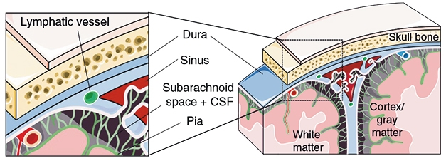
Meningeal Layers. The meninges that protect the brain comprise the dura close to the skull, the subarachnoid space bathed in cerebrospinal fluid, and the pia, which covers the gray and white matter of the brain. Shafflick separated all three and tested them for lymphoid cells. [Courtesy of Shafflick et al., Nature Neuroscience, 2021.]
Zu Hörste set out to better characterize the lymphocytes in the brain meninges. First author David Shafflick and colleagues used single-cell RNA-Seq to profile cells of the rat brain's membranes. He looked separately in the three meningeal layers: the outer dura, the middle arachnoid layer that is bathed with cerebrospinal fluid, and the inner pia that covers the brain tissue (see image above). From 26,301 total cell transcriptomes, he identified 12 major cell types among six tissues: CNS, subdural meninges, the dura, choroid plexus, CSF, and blood.
Focusing specifically on the dura, Shafflick found that it had the most diverse population of all the tissues, sporting 17 transcriptional clusters. Four subclusters of B cells (Bc) reside there. Interestingly, the Bc cluster expressed genes involved in proliferation and Nf-κB signaling, processes usually associated with B cell mobilization in the bone marrow or spleen. “This unique phenotype and expression pattern suggested dura as a potential site of homeostatic Bc residence and proliferation,” the authors wrote. Almost all the transcription patterns seen in rat dura were found in mouse dura as well, albeit less prominently.
Do these dural B cells respond to immune challenge? In mice that had experimental autoimmune encephalitis (EAE), a model of multiple sclerosis, dural B cells increased expression of MHC II, which is involved in antigen presentation, and they also upregulated ribosomal and transcription factor genes. They shut off expression of Ki67, a gene involved in proliferation. All told, it appeared that these B cells had left a homeostatic proliferation state to adopt a mature, antigen-presenting gait.
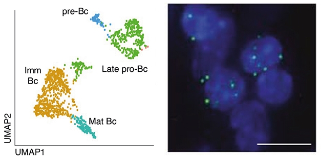
Where do these B cells come from? Unexpectedly, Shafflick found two transcriptional clusters in rat dura indicative of B cell progenitors. Dubbed pre-Bc and late pro-Bc, these clusters resembled progenitor cells that previously had been detected only in the bone marrow (see image below). Mouse dura contained similar clusters. Using flow cytometry, the authors also detected cells expressing immature B cell markers in the mouse dura. These data raise the tantalizing idea that the dura contains its own supply of B cell progenitors and does not rely on obtaining them from the bone marrow.

Dural Progenitors. Single cell RNA-Seq of the dura (left) identified transcriptional profiles in common with immature (brown) and mature (turquoise) B cells, but also with pre-B and late pro-B cell progenitors. In situ hybridization (right) identified dural cells expressing the progenitor genes dntt (red), Igll1 (white), and the common B cell marker CD19 (green). [Image courtesy Shafflick et al., Nature Neuroscience, 2021.]
Further evidence for this came from chimeric mice. The authors introduced bone marrow from a mouse that harbors the CD45.1 isoform of the cell surface protein tyrosine phosphatase into a mouse that expresses the CD45.2 isoform. All lymphocytes express CD45, and both these isoforms are equally functional, hence CD45.1 served merely to mark cells as originating from the graft. Injecting the CD45.1 bone marrow into the tail veins of CD45.2 mice after they had received a sublethal dose of radiation ensured that the host cells would be replenished with CD45.1-positive cells. Indeed, after eight weeks, about 95 percent of B cells in the lymph and skull bone marrow were replaced with donor cells; alas, in the dura, 15 percent of the B cells were still from the host. This implies either that these cells were more resistant to irradiation, or that they were replenished from within the dura, not from the injected bone marrow.
To avoid the vagaries of radiation, the authors next turned to parabiosis. They hooked up mice to a partner that ubiquitously expresses a red fluorescent protein. After seven to 10 weeks, B cells in blood, spleen, and lung were at a 1:1 ratio between host and parabiosis partner, i.e., 50 percent of the cells were read in each animal. In contrast, B cells in the host's dura were still 78 percent from the host; only 22 were red. Skull bone marrow was similarly discerning, having 34 percent red B cells. The findings are in keeping with the idea that both the dura and the skull bone marrow are replenished more slowly, or that they have their own source of B cell progenitors.
Could the skull bone marrow be supplying B cells to the dura then, as Kipnis and Colonna had previously reported? Shafflick and colleagues do not think so. They labelled B cells in the bone marrow with a dye and watched through the skull to see where these cells ended up. Even 17 days after induction of EAE, no labelled cells were found in the dura. “The skull BM thus did not likely provide large amounts of leukocytes to the dura in neuroinflammation,” conclude the authors. That’s likely to be a topic of debate and more research. —Tom Fagan
References
News Citations
Further Reading
Primary Papers
- Schafflick D, Wolbert J, Heming M, Thomas C, Hartlehnert M, Börsch AL, Ricci A, Martín-Salamanca S, Li X, Lu IN, Pawlak M, Minnerup J, Strecker JK, Seidenbecher T, Meuth SG, Hidalgo A, Liesz A, Wiendl H, Meyer Zu Horste G. Single-cell profiling of CNS border compartment leukocytes reveals that B cells and their progenitors reside in non-diseased meninges. Nat Neurosci. 2021 Sep;24(9):1225-1234. Epub 2021 Jul 12 PubMed.
Annotate
To make an annotation you must Login or Register.

Comments
Washington University in St. Louis, School of Medicine
Washington University in St Louis
This is an interesting paper that adds to recent work demonstrating that tissue-resident B cell progenitors and mature B cells reside in the dura matter, and it highlights their potential homeostatic functions. Additionally, these data add support to the idea that dural immunity may be an important site in neuroinflammatory conditions, including autoimmune diseases (Rustenhoven et al., 2021).
Interestingly, the authors suggest that B cell recruitment from local bone marrow is not a major source of infiltration—rather, their maintenance occurs predominantly via dural precursors. Given that different studies (Cugurra et al., 2021; Brioschi et al., 2021) use different approaches to answer these questions, the exact contribution from each route remains to be clarified.
References:
Rustenhoven J, Drieu A, Mamuladze T, de Lima KA, Dykstra T, Wall M, Papadopoulos Z, Kanamori M, Salvador AF, Baker W, Lemieux M, Da Mesquita S, Cugurra A, Fitzpatrick J, Sviben S, Kossina R, Bayguinov P, Townsend RR, Zhang Q, Erdmann-Gilmore P, Smirnov I, Lopes MB, Herz J, Kipnis J. Functional characterization of the dural sinuses as a neuroimmune interface. Cell. 2021 Jan 18; PubMed.
Cugurra A, Mamuladze T, Rustenhoven J, Dykstra T, Beroshvili G, Greenberg ZJ, Baker W, Papadopoulos Z, Drieu A, Blackburn S, Kanamori M, Brioschi S, Herz J, Schuettpelz LG, Colonna M, Smirnov I, Kipnis J. Skull and vertebral bone marrow are myeloid cell reservoirs for the meninges and CNS parenchyma. Science. 2021 Jun 3; PubMed.
Brioschi S, Wang WL, Peng V, Wang M, Shchukina I, Greenberg ZJ, Bando JK, Jaeger N, Czepielewski RS, Swain A, Mogilenko DA, Beatty WL, Bayguinov P, Fitzpatrick JA, Schuettpelz LG, Fronick CC, Smirnov I, Kipnis J, Shapiro VS, Wu GF, Gilfillan S, Cella M, Artyomov MN, Kleinstein SH, Colonna M. Heterogeneity of meningeal B cells reveals a lymphopoietic niche at the CNS borders. Science. 2021 Jun 3; PubMed.
Washington University School of Medicine
I wish to congratulate the authors for their exciting publication and valuable contribution. Meningeal B cells have been hiding in plain sight, and yet they have received little attention. I believe that our studies will pave the way for a fertile line of research for years to come. It is intriguing to think of the meninges as a nursery for developing immune cells, such as B cells and possibly others. I am also fascinated by B cell trafficking through the dura lymphatics. Future studies need to reveal the biological meaning of this unexpected finding.
Meningeal B cells might use the dura lymphatics as an exit route from the CNS compartment. Alternatively, this trafficking could be a highly specialized behavior of meningeal B cells to transport CNS antigens towards the cervical lymph nodes. I am sure that time will provide us with some answers. In our recent work, we provided evidence of early B cells migrating from the calvaria to the dura through a network of skull vascular channels. Here, Schafflick et al. propose that B cells progenitors may seed the dura during development and be maintained there long-term. This is certainly an intriguing hypothesis that deserves further investigation.
Make a Comment
To make a comment you must login or register.