Neurons and Cancer Cells Share Survival Tactics
Quick Links
How do neurons endure for the life of an organism? In the July 15 Science Signaling, researchers led by Mohanish Deshmukh at the University of North Carolina, Chapel Hill, report that these cells can resist insults that cause other cells to commit suicide. The researchers identified parkin-like cytoplasmic protein (PARC) as a survival factor that protects neurons by triggering the degradation of cytochrome c. Normally this respiratory chain component activates apoptotic cell death when it escapes mitochondria into the cytoplasm. Intriguingly, cancer cells also use PARC to avoid apoptosis, the authors found. “Neurons evolved this mechanism to promote their survival, and cancer cells hijacked it,” Deshmukh told Alzforum.
“The study is rigorous and extremely interesting. It provides another clue as to how neurons live for decades,” Douglas Green at St. Jude Children’s Research Hospital in Memphis, Tennessee, wrote to Alzforum.

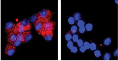
Cytochrome c (red) localizes to mitochondria in healthy neurons (left panel; nuclei in blue), but leeches into the cytoplasm if the cells lose the trophic support of growth factors. When that happens, PARC degrades the enzyme (right panel). [Image courtesy of Vivian Gama.]
Mitochondria are the energy factories of the cell. When they rupture under extreme cellular stress, free cytochrome c binds to apoptotic protease-activating factor 1 (Apaf-1), forming an “apoptosome" that then switches on caspases. These enzymes chew up cellular proteins, leading to cell death. Deshmukh and colleagues previously showed that neurons can survive even after Cyt c release—the mitochondria can recover and the cell continues to function. In part, neurons do this by inhibiting caspases. They also keep Cyt c inactive by maintaining it in a reduced state (see Potts et al., 2003; Vaughn and Deshmukh, 2008).
Deshmukh wondered what becomes of the Cyt c that floods damaged neurons. To investigate, first author Vivian Gama stressed mitochondria in cultured cells using several methods, including cell death agonists, starvation, irradiation, and the toxin staurosporine. In mouse fibroblasts, Cyt c accumulated. However, in mouse sympathetic neurons, as well as in other postmitotic cells such as cardiomyocytes, Cyt c levels were undetectable (see image above). That changed when the authors blocked the activity of the proteasome, a tube-like organelle that chews up proteins. Cyt c then built up in the cytoplasm, indicating that the proteasome normally eliminates this protein from neurons.
Proteasomes digest only proteins that are tagged with an ubiquitin peptide, which is added by an E3 ligase. To find the one that labeled Cyt c, the authors screened an siRNA library of human E3 ligases. They added the siRNAs to stressed cells to knock down each ligase in turn, and looked for an increase in Cyt c, which would indicate a failure to degrade the protein. Only knockdown of PARC pumped up Cyt c levels. PARC, which is also known as cullin-9 (CUL9), belongs to the same family as Parkin, the E3 ligase that marks damaged mitochondria for destruction and is genetically linked to Parkinson’s disease (see Feb 2010 news story; Aug 2013 news story). PARC mutations have not been tied to Parkinson’s, or Alzheimer’s, but possible associations with amyotrophic lateral sclerosis are noted on ALSGene. Intriguingly, mitochondrial dysfunction characterizes ALS (see Nov 2011 news story). Deshmukh and colleagues found that neurons and other postmitotic cells contain high levels of PARC.
Does PARC directly interact with Cyt c? The two proteins immunoprecipitated together, suggesting they formed a complex. In vitro, PARC added ubiquitin to recombinant Cyt c. In cell cultures, knockdown or deletion of PARC caused Cyt c to accumulate after mitochondrial damage in several postmitotic cell types. In addition, neurons from PARC knockout mice were more vulnerable to stress than wild-type neurons. While wild-type neurons recovered from 18 hours of growth factor deprivation, the knockout cells died. Conversely, overexpressing PARC in various dividing cells lowered Cyt c levels and improved survival after stress.
What about cancer cells, which also resist apoptosis? Neuroblastoma and glioblastoma cell lines likewise kept Cyt c levels low under stress, the authors saw. While PARC levels were not particularly high in these lines, blocking the enzyme in stressed cells caused Cyt c to build up, and more cells died. The results suggest that cancer cells use the same mechanism as neurons to dodge death, Deshmukh said. In ongoing work, he is examining whether brain cancers are more sensitive to chemotherapy in PARC knockout mice compared to wild-type.
Some epidemiological studies have found an inverse correlation between cancer and neurodegeneration, which has been attributed to genetic differences in cell survival pathways (see Dec 2009 news story; Oct 2010 conference story). “The data suggest that if you are genetically more resistant to apoptosis, your neurons will be better protected, but you may also be predisposed to getting cancer,” Deshmukh said.
Commentators hailed the findings as an advance in understanding cell death pathways. “This study’s findings complement the wealth of data linking regulation of cell death and survival by the ubiquitin-proteasome system. Moreover, the authors identify Cyt c as the first known substrate of PARC/CUL9 ubiquitin ligase,” Jonathan Lopez and Stephen Tait of the University of Glasgow, Scotland, wrote in an accompanying commentary.
“The paper is an exciting, elegant piece of work,” said Miratul Muqit at the University of Dundee, Scotland. “The data suggest that inhibition of PARC could be a tractable therapeutic target for cancer.” Green noted that PARC also squelches apoptosis by anchoring the tumor suppressor p53 in the cytoplasm, preventing it from traveling to the nucleus and triggering cell death. For that reason, pharmaceutical companies already want to inhibit PARC in cancer cells. “These data provide another reason to do so,” Green added. Drug companies have more experience at inhibiting kinases than the ubiquitin system, but Muqit noted that interest in the latter target is growing.
It is less clear how to target PARC to improve neuronal survival. Muqit pointed out that PARC contains a second active domain in addition to its E3 ligase. This is a cullin domain, which acts as a scaffold to recruit E2 ligases that can also ubiquitinate proteins. Which activity mediates the degradation of Cyt c? Further work could nail this down and help researchers devise better ways to intervene therapeutically, said Muqit.
Meanwhile, Deshmukh thinks PARC might be useful for studying Parkinson’s. Parkin knockouts have been disappointing as PD models because they do not recapitulate all the symptoms of the disease. However, Parkin and PARC may act in a complementary fashion, since the former helps destroy damaged mitochondria and the latter targets a mitochondrial protein. “We think that knocking out both could create a better model, because then neurons would have no capability for dealing with damaged mitochondria,” he said.—Madolyn Bowman Rogers.
References
News Citations
- Abnormal Mitochondrial Dynamics—Early Event in AD, PD?
- Parkinsonism-linked Protein Binds Parkin and Pink1, Drives Mitophagy
- Mutant Meddling in Mitochondria Partly Mimics ALS Pathology
- Research Brief: Epidemiological Study Links Cancer, AD
- Two Faces of Evil: Cancer and Neurodegeneration
Paper Citations
- Potts PR, Singh S, Knezek M, Thompson CB, Deshmukh M. Critical function of endogenous XIAP in regulating caspase activation during sympathetic neuronal apoptosis. J Cell Biol. 2003 Nov 24;163(4):789-99. Epub 2003 Nov 17 PubMed.
- Vaughn AE, Deshmukh M. Glucose metabolism inhibits apoptosis in neurons and cancer cells by redox inactivation of cytochrome c. Nat Cell Biol. 2008 Dec;10(12):1477-83. Epub 2008 Nov 23 PubMed.
External Citations
Further Reading
Primary Papers
- Gama V, Swahari V, Schafer J, Kole AJ, Evans A, Huang Y, Cliffe A, Golitz B, Sciaky N, Pei XH, Xiong Y, Deshmukh M. The E3 ligase PARC mediates the degradation of cytosolic cytochrome c to promote survival in neurons and cancer cells. Sci Signal. 2014 Jul 15;7(334):ra67. PubMed.
- Lopez J, Tait SW. Killing the Killer: PARC/CUL9 promotes cell survival by destroying cytochrome C. Sci Signal. 2014 Jul 15;7(334):pe17. PubMed.
Annotate
To make an annotation you must Login or Register.

Comments
No Available Comments
Make a Comment
To make a comment you must login or register.