Digital Microscopy Neuropathology Collection
The digital microscopy platform for neurodegenerative diseases is being developed by Jochen Herms and Sigrun Roeber at the Center for Neuropathology and Prion Research of Ludwig Maximilian University, Munich, Germany. The collection provides high-resolution histological specimens characteristic of such diseases for the purposes of learning and teaching. The site is freely available for registered users; to register, click here.
The collection includes images of brain tissue from healthy individuals and from people who had Alzheimer’s disease, Parkinson's disease, Frontotemporal Lobar Degeneration, Amyotrophic Lateral Sclerosis, different types of prion disease, or a range of other, rare neurodegenerative disorders. As the curators expand and develop the collection, they are inviting user feedback and suggestions on what else to include via their email addresses on the platform.
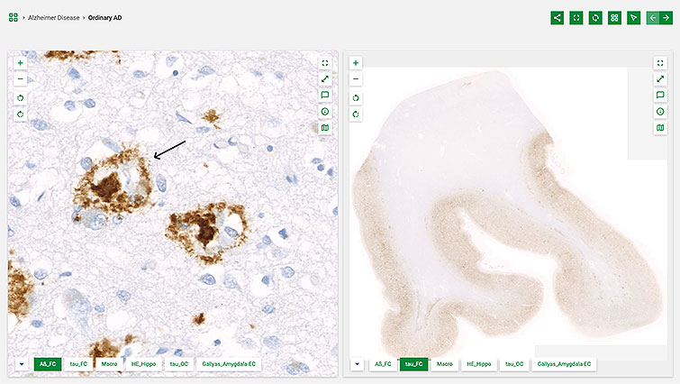
Navigation of the collection is intuitive. Some helpful hints: after selecting a disease from the homepage, users can click one of its example cases and examine multiple images from each case. Some icons enable zooming in, out, rotating, or expanding the size of the images. Others are explained via pdf. Additional navigation aids include:
 Annotation icon. Opens a list describing features of each image. Selecting a particular feature highlights it in the image with an arrow.
 Information icon. Provides details about the diagram.
 Overview map icon. Displays the relative position of the image on the gross anatomy slide.