
Are Artery Exosomes to Blame for Vascular Cognitive Impairment?
Quick Links
Atherosclerotic arteries aren’t just a problem for the heart—they’re bad news for the brain, too. Now, scientists led by Wei Wang, Dai-Shi Tian, and Chuan Qin of Huazhong University of Science and Technology in Wuhan, China, offer a new explanation. They report that arterial plaques flood the bloodstream with brain-invading exosomes that rile up microglia and damage white matter. The likely source of this onslaught? Macrophages stuffed with fat—aka foam cells. Published May 8 in Cell Metabolism, the findings evoke a new mechanism by which hardened vessels lead to cognitive decline.
- In mice, a high-fat diet triggers atherosclerotic plaques in blood vessels.
- Macrophages in the plaques release exosomes.
- These activate microglia in the brain, worsening white matter damage and memory.
“This is an interesting paper,” said Robert Rissman of the University of Southern California, Los Angeles. “Not only because it implicates specific extracellular vesicles released during atherosclerosis in cognitive impairment, but also because it highlights how peripheral inflammation might directly influence the brain.”
Tsuneya Ikezu, Mayo Clinic, Jacksonville, Florida, found the paper fascinating. “The work compellingly demonstrates the migration of plasma extracellular vesicles (EVs) into the CNS, where they are predominantly taken up by microglia,” he wrote to Alzforum. However, Ikezu was unconvinced this leads to white-matter damage. “Demonstrating a microglia-dependent mechanism would significantly strengthen the conclusions,” he added (comment below).
Atherosclerosis—the buildup of fatty gunk in artery walls—is linked to vascular cognitive impairment, but scientists have struggled to pin down how these fatty lesions compromise brain health. Enter exosomes. Wrapped into a lipid bilayer, these tiny messengers ferry a variety of biological molecules over long distances. Atherosclerotic vessels release large quantities of circulating exosomes into the blood (for a review see Wang et al., 2021), hence researchers suspect that these stealthy couriers may be the missing link.
To test this idea, first authors Hang Zhang, Luo-Qi Zhou, Sheng Yang, and colleagues devised a two-stage mouse model. First, they harvested and purified exosomes from either atherosclerotic or healthy mice. Then, they infused these vesicles into a second cohort with a surgically induced vascular cognitive impairment. This design enabled them to measure the vesicles’ effects directly, independent of other complications tied to atherosclerosis.
Before collecting exosomes, Zhang and colleagues put young ApoE-knockout mice on a high-fat, Western-style diet. ApoE is essential for clearing lipoproteins from the blood; without it, mice developed lipid-rich deposits that caked their arteries. After 14 weeks, the scientists collected blood plasma and found that these mice had churned out far more exosomes than mice on a healthier diet (image below).
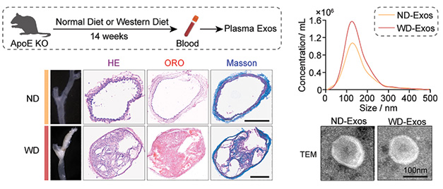
Blocked Blood Vessels, Extra Exosomes. ApoE-knockout mice fed a high-fat diet accumulate fatty deposits in their arteries, as revealed by H&E, Oil Red O (lipids), and Masson’s trichrome staining (fibrous tissue) of plaque cross-sections (bottom). They produce more circulating exosomes than do mice on normal chow (right). [Courtesy of Zhang et al., Cell Metabolism, 2025.]
Next came the exosome recipients. To model vascular cognitive impairment, the scientists wrapped tiny coils around the carotid arteries supplying the brains of wild-type mice, restricting blood flow and triggering white-matter damage. One week later, they injected these mice with exosomes—either from the Western-diet–fed mice or from healthy controls. They gave 10 injections over 28 days, then examined the brains for myelin damage. Exosomes from atherosclerotic mice had exacerbated white-matter loss beyond the effect of the arterial occlusion alone, while exosomes from healthy mice had no such impact. Electron microscopy told the same story, showing frayed, loosened myelin sheaths (image below).
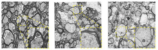
Messed-Up Myelin. Transmission electron microscopy shows thinning of myelin around axons of the corpus callosum. Control mice have normal myelination (left). Carotid artery stenosis induces myelin loss (middle). Exosomes from mice fed a fatty diet worsen the effect of the stenosis (right). [Courtesy of Zhang et al., Cell Metabolism, 2025.]
This took a toll on memory. In the eight-arm maze, where mice typically explore each arm just once to find a treat, those injected with Western-diet exosomes repeatedly revisited the same arms, unable to remember they'd been there before.
To learn where these vesicles end up in the brain, the researchers labeled them with the lipophilic dye PKH26, injected them, and tracked them. Nearly half of the tagged vesicles turned up inside microglia. Surveying microglia in exosome-treated mice revealed telltale signs of activation: Their numbers in the brain swelled, they bulked up, and levels of inflammatory and disease-associated markers rose.
Might these microglia be the ones torching white matter? To find out, scientists fed mice a diet laced with PLX5622, a CSF1R inhibitor that depletes resident microglia. Without microglia, the Western-diet exosomes lost their pathogenic punch. Demyelination eased (image below) and maze performance improved compared to mice with intact microglia.
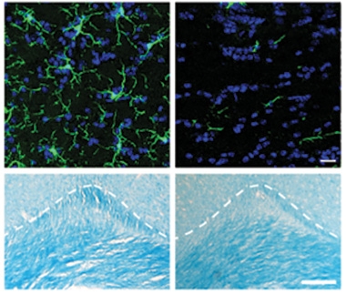
No Microglia, No Problem. In mice given atherosclerotic exosomes (left), microglia (top) become activated (green) and myelin (blue) degrades (bottom). Depleting microglia with PLX5622 (right) preserves white-matter integrity. [Courtesy of Zhang et al., Cell Metabolism, 2025.]
Where do these poison-pill exosomes come from? To trace the perp, scientists turned to single-nucleus RNA sequencing of cells in human carotid plaques. They lined up the suspects—smooth muscle cells, endothelial cells, T cells, B cells, myeloid cells. Only the latter produced transcripts that support exosome-assembly. After further interrogation, one myeloid cell subset stood out: hello, foam cells. These macrophages were the most transcriptionally primed to launch an exosomal assault.
To investigate the effects of foam cell exosomes, researchers generated them in a dish. They cultured bone marrow cells from mice, differentiated them into macrophages, and transformed these into foam cells by treating them with oxidized LDL for 24 hours. Then they took exosomes from the culture media, let them loose on mouse primary microglia, and measured changes in the cells’ transcriptomes (image at right). Compared to exosomes from regular macrophages, those from foam cells sparked a surge in proinflammatory gene expression and cytokine release—pointing squarely to these cells as potent microglial agitators.
What do these exosomes carry that so easily riles microglia? One clue came from the microglial transcriptome. The biggest change was in genes that can be silenced by micro RNAs. Twenty miRNAs were abundant in the exosomes. Of these, six were predicted to regulate genes that were suppressed in the microglia. Pathway analysis suggested these six miRNA/mRNA pairs would hamstring phagosomes, myelination, and an oxidative stress response driven by the transcription factor Nrf2. The latter stood out because Nrf2 is a master regulator of antioxidant defenses and controls the expression of genes that neutralize reactive oxygen species and maintain mitochondrial health. In microglia exposed to foam-cell–derived exosomes, Nrf2 activity fell, as did expression of the glucose transporter GLUT1. Glucose metabolism tanked, oxidative stress ramped up, and mitochondria damage ensued. Bumping up expression of Nrf2 in mice with an AAV vector prevented microglial activation and white-matter injury when the animals were exposed to atherosclerotic exosomes.
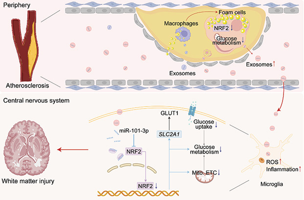
Plaque Problems. Lipid-laden foam cells churn out exosomes loaded with inflammatory cargo (top). These vesicles enter the bloodstream, head to the brain, and are gobbled up by microglia (bottom). Once inside, they spark inflammation and reduce antioxidant defenses by suppressing Nrf2. The end result? Fraying white matter and faltering memory. [Courtesy of Zhang et al., Cell Metabolism, 2025.]
The findings address a conundrum that has puzzled scientists for years, namely, that reduced cerebral blood flow alone cannot explain cognitive impairment seen in people with atherosclerosis. Indeed, the authors confirmed this with data from the U.K. Biobank. They found that among 1,630 participants with carotid artery thickening and white-matter hyperintensities on MRI scans, arterial plaque correlated with white-matter damage, even after accounting for reduced cerebral blood flow and other common risk factors.
Taken together, the researchers propose a model in which exosomes released from foam cells travel to the brain, push microglia into a pro-inflammatory state, and leave white matter in tatters. This chain of events, they suggest, helps precipitate cognitive decline. One caveat? The authors relied on carotid artery stenosis. Large vessel atherosclerosis, such as in carotids or vertebrobasilar arteries, has a very weak relationship to cognition, scientists told Alzforum. For the proposed mechanism to be potentially meaningful, it would also have to occur in arteriolosclerosis, i.e., the smaller penetrating arterioles.—George R. Heaton
George Heaton is a freelance writer in Durham, North Carolina.
References
Paper Citations
- Wang C, Li Z, Liu Y, Yuan L. Exosomes in atherosclerosis: performers, bystanders, biomarkers, and therapeutic targets. Theranostics. 2021;11(8):3996-4010. Epub 2021 Feb 15 PubMed.
Further Reading
Papers
- Wang C, Li Z, Liu Y, Yuan L. Exosomes in atherosclerosis: performers, bystanders, biomarkers, and therapeutic targets. Theranostics. 2021;11(8):3996-4010. Epub 2021 Feb 15 PubMed.
Primary Papers
- Zhang H, Zhou LQ, Yang S, Dong MH, Chen L, Lu YL, Zhang LY, Zhang L, Chu YH, Xu LL, Pang XW, Zhu LF, Xu T, Yong TY, Wang W, Tian DS, Qin C. The foam cell-derived exosomes exacerbate ischemic white matter injury via transmitting metabolic defects to microglia. Cell Metab. 2025 Aug 5;37(8):1636-1654.e10. Epub 2025 May 8 PubMed.
Annotate
To make an annotation you must Login or Register.
Comments
Alzheimer's Therapeutic Research Institute, University of Southern California
This is an interesting paper. Not only because it implicates specific extracellular vesicles released during atherosclerosis in cognitive impairment, but also because it highlights how peripheral inflammation might directly influence the brain. That these specific EVs are involved is a critical finding, but, more generally, the paper also provides insight into possible new links between peripheral inflammatory pathways and the brain. What isn't particularly clear is whether there is a specific cargo that causes this effect, or it's the EV itself or something else. There is some insight of pathways that may be involved.
Another interesting aspect is the use of the word, exosome. The ADRD field has largely departed from this term as it's been very difficult to specifically identify a specific species of vesicles and to completely exclude others. I would caution the authors regarding the use of this term as it's likely that exosomes are only one subtype of EV that may be involved in this effect.
We also do not understand much about the trafficking of EVs and how they develop the cargos and external signals/markers that we find them with. For example, these foam-cell derived EVs may be released in a specific state with specific cargos and external markers, but whether they retain these markers or cargos when they hit their final target is unknown. That is, it is unclear that the original exosome/EV is the actual mechanistic link in the effects seen.
Mayo Clinic Florida
This fascinating study on the impact of foam-cell-derived exosomes on ischemic CNS injury compellingly demonstrates the migration of plasma extracellular vesicles (EVs) into the CNS, where they are predominantly taken up by microglia. That said, I would advise caution in interpreting these findings, as intravenously administered EVs typically show limited CNS penetration and are largely sequestered by the liver. Additionally, the role of microglia in EV-mediated white-matter pathology remains unclear, particularly in the context of PLX5622 treatment. Demonstrating a microglia-dependent mechanism would significantly strengthen the conclusions.
The molecular analysis of how EV-associated hsa-miR-101-3p disrupts NRF2 signaling is impressively thorough, incorporating both in vitro and in vivo approaches. However, EVs also carry bioactive lipids and proteins that may contribute to disease processes, which were not explored in this study.
Moreover, the use of AAV for microglia-specific gene delivery raises some concerns, as its efficiency in targeting microglia is not well established. The manuscript would benefit from more detailed information regarding the AAV serotype used, transduction efficiency, and cell-type specificity.
Nonetheless, the concept that inhibiting EV secretion from foam cells could offer therapeutic potential for atherosclerosis-related CNS disorders is intriguing and opens up promising avenues for future research.
Newcastle University and University of Nairobi, Kenya
It is estimated that carotid artery disease causes 10-15 percent of ischemic strokes. Stroke injury is most often accompanied by diffuse white-matter disease. This is characterized by the “white caps” in the periventricular region and white-matter hyperintensities (WMH) in the deep white matter seen upon MRI. Various mechanisms have been suggested to explain how cerebral white-matter disease of vascular origin may occur and progress. While the association between carotid artery disease and stroke is known in that carotid intimal-media thickness (CIMT) is acknowledged as an early and measurable indication for incident stroke and cardiovascular events, the association with white-matter disease has been less well established.
In a proportion of dementia patients, walls of internal carotid arteries may develop atherosclerotic plaques to cause near enough 100 percent stenosis to reduce blood flow to the brain. Moderate to severe stenosis may lead to cerebral hypoperfusion, contributing to white-matter changes or WMH without other influences, and these are found not only in VCI and vascular dementia, but also in neurodegenerative dementias such as Alzheimer’s disease.
Atherosclerotic plaques typically accumulate fragmented smooth muscle cells, macrophages, lymphocytes, lipids, extracellular matrix proteins and foam cells. Classically foam cells may have two lineages: Some are macrophages derived from monocytes, which adhere to and penetrate the endothelium, and some are from medial smooth muscle cells, which proliferate and penetrate the intima. However, cellular elements, which accumulate in the atheromas, could rupture and release contents to potentially instigate other damage to the cerebral endothelium and brain parenchyma if these elements travel upstream to the brain.
In this cutting-edge study, Zhang et al., using a large dataset from the U.K. Biobank, first showed that there was an independent association between carotid stenosis (CIMT as a proxy for atherosclerotic disease) and WMH. Then the authors cleverly elucidated that exosomes from macrophage-derived foam cells originating from atherosclerotic plaques (results from experimental mice models and human carotid atheromas) essentially exacerbate ischemic white-matter injury. These foam cells transmit glucose metabolic defects to microglia via an exosomal miRNA (miR-101-3p), which they identified as the key molecule using single-cell transcriptomic studies. They found that the old friend Nrf2 transcriptionally activates microglia associated GLUT1 (Slc2a1) to enhance glucose metabolism.
Of course, this study does not address how the identified mechanisms, including glucose metabolic effects, could influence oligodendrocytes or other cells within the gliovascular unit to affect myelination or the wiring of the brain. Notwithstanding, this remarkable study has implications for several dementias, including AD, which are not necessarily immune to coronary artery, carotid artery or intracranial atherosclerosis. The study clearly advances the field, although it is difficult to appreciate to what degree exosomes derived from foam cells contribute to white-matter injury as it would depend on the extent of arterial atherosclerosis, which exponentially increases with age.
Make a Comment
To make a comment you must login or register.