
Blood Tests Forecast Dementia in Down’s Syndrome
Quick Links
Up-and-coming Alzheimer’s blood tests may soon be used for people with Down’s syndrome. In a longitudinal biomarker study of adults with three copies of chromosome 21, investigators from the Alzheimer’s Biomarker Consortium–Down Syndrome (ABC-DS) report that elevated plasma p-tau217 signals advancing amyloid and tau pathology and progression to dementia. Published June 12 in The Lancet Neurology, the study, led by Oskar Hansson at Lund University, Sweden, also found that high plasma GFAP foreshadowed amyloid accumulation. Their findings echo an independent study published June 8 in the Cell Press journal Med by Madhav Thambisetty and colleagues at the National Institutes of Health, Baltimore. It shows that, in otherwise healthy people, blood GFAP begins to climb a decade before symptoms of Alzheimer’s disease appear.
- In Down’s syndrome, plasma p-tau217 ticks up.
- It predicts amyloid and tau pathology.
- High plasma GFAP also signals amyloid buildup.
“Plasma p-tau217 provides a noninvasive, highly scalable, and widely accessible method for identifying individuals at risk for AD progression,” said Michael Rafii of the University of Southern California, Los Angeles. “These findings further validate the use of pathological biomarkers—rather than relying solely on clinical symptoms—for diagnosing Alzheimer’s disease in Down’s syndrome,” he added (comment below).
Many scientists view Down’s syndrome as a genetic form of Alzheimer’s because the amyloid precursor protein (APP) gene lies on chromosome 21. The extra dose of APP in DS causes amyloid to amass in the brain, putting everyone one with trisomy 21 at high risk for Alzheimer’s dementia. When plasma p-tau217 burst onto the scene as a leading Alzheimer’s biomarker, it motivated scientists to see whether it would identify AD in people with Down’s, too. That hunch proved correct. In 2022, ABC-DS investigators reported that p-tau217 in the blood of people with DS mirrored amyloid plaques and tau tangles in the brain (Janelidze et al., 2022). Now they ask: Could blood biomarkers forecast future pathology—and the dementia that follows?
To investigate, ABC-DS researchers analyzed data from 258 adults with Down’s who had been recruited from seven sites across the U.S. and U.K between 2016 and 2019 (image below). The average age was 45; 53 percent were women. Every 16 months, participants sat for a cognitive checkup and rolled up their sleeves for blood samples. For this paper, first authors Shorena Janelidze, Lyduine Collij, and colleagues measured five biomarker candidates in these samples—p-tau217, GFAP, Aβ42/40, neurofilament light chain (NfL), and total tau. Only those participants who returned for at least one follow-up visit were included in this analysis. When ABC-DS began, 195 showed no signs of dementia, 34 had mild cognitive impairment, and 23 met criteria for dementia. A subset also had their brains scanned to track pathology: 106 logged two or more amyloid-PET scans, and 89 had sequential tau-PET scans.
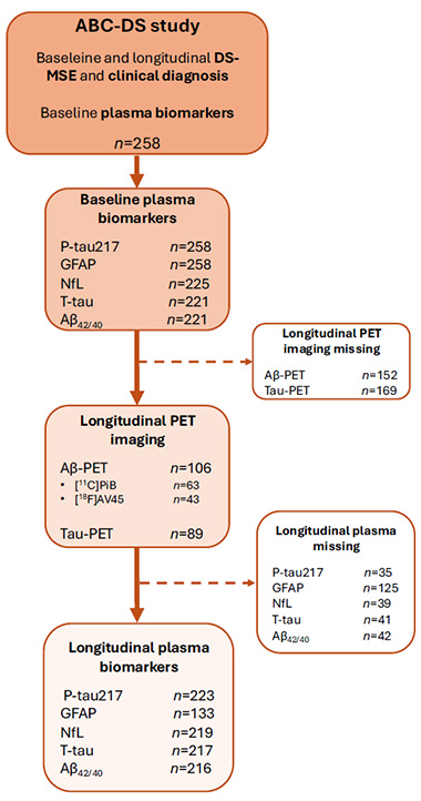
Blood and Brain. Flow chart of participant recruitment and retention, showing follow-up for cognitive testing, blood biomarker sampling, and PET imaging. [Courtesy of Janelidze et al., The Lancet Neurology, 2025.]
With all the data gathered and the number-crunching done, what did Janelidze and Collij find? At baseline, higher p-tau217, GFAP, NfL, and total tau were each individually linked to a greater risk of dementia, as was a lower Aβ42/40 ratio. Of these, p-tau217 levels stood out as the most reliable. So much so, in fact, that when all the markers were lumped together in the same analysis, it was the only one that consistently pointed to who was most likely to develop dementia.
In AD, p-tau217 has emerged as a robust marker of amyloid pathology, likely reflecting tau phosphorylation that occurs downstream of amyloid toxicity (Dec 2022 conference news; Aug 2024 conference news).
In the Down’s cohort, plasma p-tau217 also tracked with tangles. In participants who had sequential tau PET scans, elevated p-tau217 predicted a rise in tangles (image below). High plasma p-tau217 was especially ominous in participants who were already amyloid-positive, boosting the chances that more tangles would show up later, as is the case for non-DS Alzheimer’s (Dec 2021 news). This is in keeping with p-tau217 being a better marker for the presence of amyloid pathology. As for amyloid itself, PET scans revealed that higher baseline levels of p-tau217 and the astrocytic protein GFAP signaled more plaques at follow-up.
“The associations between biomarker changes and changes in cognition, Aβ-PET, or tau-PET in people with Down's syndrome are very similar to the associations seen in Alzheimer’s disease in general,” Charlotte Teunissen and Flora Duits, Amsterdam University Medical Center, Netherlands, wrote in an accompanying editorial. “This finding suggests not only similar pathophysiology, but also similar potential uses of these biomarkers for diagnosis and treatment response monitoring,” they added.
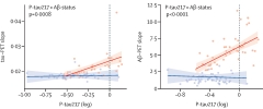
Predicting PET. In people with Down’s syndrome (Aβ-positive, blue; Aβ-negative, red), plasma p-tau217 correlated with change in tau PET (left) and amyloid PET scans (right). [Courtesy of Janelidze et al., Lancet Neurology, 2025.]
Janelidze and colleagues weren’t the only ones to notice the GFAP alarm bell. Thambisetty’s group analyzed plasma biomarkers in participants from the Baltimore Longitudinal Study of Aging, a 67-year-old cohort study tracking age-related changes. For this paper, first author Vijay Varma identified participants who were cognitively unimpaired but later developed mild cognitive impairment or dementia. He matched a total of 158 of them with participants who remained cognitively sharp and shared the same sex, race, approximate age, and visit year.
Compared to controls, baseline plasma GFAP ran higher in people who developed AD, with differences appearing a decade prior to diagnosis (image below). No other plasma markers, including Aβ42/40, p-tau181, or p-tau231, differed between converters and controls prior to Alzheimer’s onset. P-tau217 was not measured in this study.
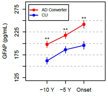
Mind the GFAP. People who developed AD had higher plasma GFAP 10 or 5 years before their symptoms started, or at the time of onset, than did those who stayed cognitively healthy. [Courtesy of Varma et al., Med, 2025.]
Although PET imaging data for amyloid or tau was not collected in this study, which has been running since 1958, Varma and colleagues examined neuropathology in a separate group of participants at postmortem. Consistent with their previous findings, plasma GFAP levels, measured from a blood draw taken a few years before death, were highest in those with Alzheimer’s compared to controls. GFAP also tracked with the severity of amyloid pathology, as assessed by CERAD scores, and with tau pathology, as measured by Braak stage.
“This study adds to a growing body of evidence suggesting that astrocytic reactivity may act as an upstream trigger in AD pathogenesis,” said Tharick Pascoal of the University of Pittsburgh. “Recognizing this distinction is critical, as it strengthens the rationale for targeting pathological astrocyte reactivity,” he added.
Tracking GFAP levels in the blood could also help researchers determine whether an experimental Alzheimer’s treatment is slowing or altering the course of the disease, Thambisetty and colleagues suggest.
Teunissen and Duits noted that individual trajectories of biomarkers and cognition in people with Down's syndrome need further study to help define when to begin Aβ-targeting therapies, much like how inclusion criteria have been set for preclinical Alzheimer’s trials such as TRAILBLAZER-ALZ3 (NCT05026866).
Hansson and ABC-DS scientists propose that p-tau217, perhaps in combination with GFAP, could be useful in prognostic evaluations, and in clinical practice and trials involving people with trisomy 21. “Our study highlights the power of team science,” Elizabeth Head, Ben Handen, Mark Mapstone, and Brad Christian—all leaders of the consortium—told Alzforum (comment below). “We hope studies like this will create a thriving clinical trial ecosystem for people with Down’s syndrome.”—George R. Heaton
George Heaton is a freelance writer in Durham, North Carolina.
References
Mutations Citations
News Citations
- A Tau Blood Test Tracks With Alzheimer's Neuropathology
- Are Alzheimer’s Blood Tests Ready for Primary Care?
- In Preclinical Alzheimer's, p-tau217 in Blood Best Predicts Tangles
Paper Citations
- Janelidze S, Christian BT, Price J, Laymon C, Schupf N, Klunk WE, Lott I, Silverman W, Rosas HD, Zaman S, Mapstone M, Lai F, Ances BM, Handen BL, Hansson O. Detection of Brain Tau Pathology in Down Syndrome Using Plasma Biomarkers. JAMA Neurol. 2022 Aug 1;79(8):797-807. PubMed.
External Citations
Further Reading
Papers
- Janelidze S, Christian BT, Price J, Laymon C, Schupf N, Klunk WE, Lott I, Silverman W, Rosas HD, Zaman S, Mapstone M, Lai F, Ances BM, Handen BL, Hansson O. Detection of Brain Tau Pathology in Down Syndrome Using Plasma Biomarkers. JAMA Neurol. 2022 Aug 1;79(8):797-807. PubMed.
Primary Papers
- Janelidze S, Collij LE, Mattsson-Carlgren N, Antill A, Laymon CM, Lott I, Rosas HD, Minhas DS, Luo W, Zaman S, Alzheimer's Biomarker Consortium–Down Syndrome investigators, Mapstone M, Head E, Lai F, Hartley SL, Ances BM, Krinsky-McHale SJ, Lee JH, Ossenkoppele R, Christian BT, Handen BL, Hansson O. Prediction of amyloid and tau brain deposition and cognitive decline in people with Down syndrome using plasma biomarkers: a longitudinal cohort study. Lancet Neurol. 2025 Jul;24(7):591-600. PubMed.
- Varma VR, An Y, Kac PR, Bilgel M, Moghekar A, Loeffler T, Amschl D, Daurer M, Prokesch M, Troncoso J, Blennow K, Zetterberg H, Ashton NJ, Ferrucci L, Resnick SM, Thambisetty M. Longitudinal progression of blood biomarkers reveals a key role of reactive astrocytosis in preclinical Alzheimer's disease. Med. 2025 Sep 12;6(9):100724. Epub 2025 Jun 9 PubMed.
- Teunissen C, Duits F. Plasma biomarkers for diagnosis and prognosis in Down syndrome-related Alzheimer's disease. Lancet Neurol. 2025 Jul;24(7):561-562. PubMed.
Annotate
To make an annotation you must Login or Register.
Comments
University of Southern California Keck School of Mediicine
The Janelidze et al. study reinforces plasma p-tau217 as a highly reliable and predictive biomarker for amyloid accumulation, tau pathology, and now progression to dementia in individuals with Down’s syndrome. These findings further validate the use of pathological biomarkers—rather than relying solely on clinical symptoms—for diagnosing Alzheimer’s disease in DS, a genetic form of AD, much like how biomarker evidence is standard in diagnosing cancer and cardiovascular disease.
Plasma p-tau217 provides a noninvasive, highly scalable, and widely accessible method for identifying individuals at risk for AD progression. It is already being implemented across all ACTC-DS affiliated studies.
University of Pittsburgh
Varma and Thambisetty conducted a nice translational study that combined human longitudinal data, postmortem brain tissue, and a transgenic mouse model of AD. They demonstrated that plasma GFAP levels begin to increase a decade before the onset of cognitive symptoms, and that they are predictive of clinical progression. Elevated GFAP was associated with increased AD neuropathological burden and correlated with cortical GFAP immunoreactivity in mice, supporting its utility as a peripheral indicator of brain astrocytosis in AD.
This study adds to a growing body of evidence suggesting that astrocytic reactivity may act as an upstream trigger in AD pathogenesis rather than a downstream consequence of neurodegeneration. Recognizing this distinction is critical, as it strengthens the rationale for targeting pathological astrocyte reactivity as a modifiable mechanism in disease progression. On the other hand, if astrocytic reactivity were merely an epiphenomenon, therapeutic strategies targeting glial pathways would lack strong biological justification. These findings are in line with recent trends in Alzheimer's clinical trials, where neuroinflammation has become the most frequently targeted biology in ongoing Phase 2 trials.
VU University Medical Center
The results of Thambisetty’s group showing elevated GFAP levels a decade before clinical conversion to AD extend our findings in the DIAN cohort (Chatterjee et al., 2023), as well as our study in the Netherlands Twin Registry, a population-based cohort (den Braber et al., 2023). Varma and colleagues made their observations in a slightly older population (average age 65 at baseline versus 58 in the twin registry). In our study, plasma GFAP, p-tau181, and the Aβ42/40 ratio changed in those who tested positive for amyloid 10 years later (AUC for GFAP 0.84), while still not cognitively impaired.
Remarkably, in both Varma et al. and den Braber et al., the lines do not intersect at 10 years before onset in sporadic patients, which may suggest that the starting point of the change is even earlier. Alternatively, it could simply be that elevated GFAP is a trait rather than a state marker, which is further supported by the high, 60 percent, heritability of plasma GFAP levels (Rousset et al., 2025). This hypothesis of course remains to be tested further.
But what is the possible tissue origin of GFAP? The results of this study do not give a clear answer to this question. Astrocyte reactivity in the 5xFAD model was present at month 3, amyloid deposition started between months 3 and 7, but plasma levels changed only at month 7, and only at trend level. This suggests that the relation between elevated plasma GFAP and either GFAP expression in brain tissue, or levels of amyloid deposition are not very strong, This suggests that research should focus on identification of earlier astrocyte markers, since astrocytes play a relevant role in AD pathology.
References:
Chatterjee P, Vermunt L, Gordon BA, Pedrini S, Boonkamp L, Armstrong NJ, Xiong C, Singh AK, Li Y, Sohrabi HR, Taddei K, Molloy M, Benzinger TL, Morris JC, Karch C, Berman S, Chhatwal J, Cruchaga C, Graff-Radford NR, Day GS, Farlow M, Fox N, Goate A, Hassenstab J, Lee JH, Levin J, McDade E, Mori H, Perrin R, Sanchez-Valle R, Schofield PR, Levey A, Jucker M, Masters CL, Fagan AM, Bateman RJ, Martins RN, Teunissen C, and the Dominantly Inherited Alzheimer Network. Plasma glial fibrillary acidic protein in autosomal dominant Alzheimer's disease: Associations with Aβ-PET, neurodegeneration, and cognition. Alzheimers Dement. 2022 Dec 28; PubMed.
den Braber A, Verberk IM, Tomassen J, den Dulk B, Stoops E, Dage JL, Collij LE, Barkhof F, Willemsen G, Nivard MG, van Berckel BN, Scheltens P, Visser PJ, de Geus EJ, Teunissen CE. Plasma biomarkers predict amyloid pathology in cognitively normal monozygotic twins after 10 years. Brain Commun. 2023;5(1):fcad024. Epub 2023 Feb 4 PubMed.
Rousset RZ, den Braber A, Verberk IM, Boonkamp L, Wilson DH, Ligthart L, Teunissen CE, de Geus EJ. Heritability of Alzheimer's disease plasma biomarkers: A nuclear twin family design. Alzheimers Dement. 2025 Jan;21(1):e14269. Epub 2024 Nov 26 PubMed.
University of North Texas Health Science Center
Janelidze et al. is a very nice paper looking at plasma biomarkers of Alzheimer’s disease in the incredibly valuable Alzheimer’s Biomarker Consortium–Down Syndrome (ABC-DS). I am part of the ABC-DS, and assays other than for p-tau217 were run on the Quanterix systems in my lab.
There is a growing body of literature supporting the notion that several blood markers, particularly in combination, would have a range of context of use (COU) for AD. However, less work has been done specifically within this at-risk population. The Alzheimer’s Clinical Trials Consortium (ACTC)-DS at the University of Southern California is an infrastructure specifically designed to advance clinical trials among individuals with Down’s syndrome, and ABC-DS is working closely with ACTC-DS on these trials.
Prior work out of ABC-DS has shown that markers of amyloid, tau, inflammation, and others have very strong potential for use, including total tau, and NfL as shown in this current work. The current work highlighting that ptau-217, GFAP, NfL, and total tau have potential use among individuals with Down’s syndrome is a great addition to the science.
Given recent blood tests that implement p-tau217 with Aβ42 for detecting possible brain amyloid among those with cognitive impairment, these new results suggest that similar combined markers may be possible for DS. For example, can a ratio, or algorithm, that includes p-tau217 or GFAP (or possibly NfL) be utilized to create a risk score for change in amyloid PET over time for a possible surrogate outcome in a clinical trial? Can p-tau217 be combined with GFAP, NfL, and total tau to create a risk score with a 90 percent probability of cognitive decline over a given time period, e.g., two years, to selectively enroll only those patients most likely to experience decline for a trial to demonstrate stronger effect sizes?
This work not only leverages a significant amount of prior work from ABC-DS, and international collaborations, but also extends it and helps set the stage for the next steps in advancing clinical trials for this specific population. My congratulations to the team, the ABC-DS, and the participants for this wonderful work.
University of Kentucky
University of California, Irvine
University of Wisconsin-Madison
We were delighted to collaborate with Shorena Janelidze and Oskar Hansson at Lund University, and colleagues, on this landmark paper, describing the power of plasma p-tau217 in helping predict cognitive decline in people with Down’s syndrome. This exciting project stemmed from an initial meeting with the Alzheimer Biomarker Consortium—Down’s Syndrome (ABC-DS) led by principal investigators Brad Christian and Ben Handen, at the Human Amyloid Imaging meeting in 2022. From that, a paper describing cross-sectional predictive outcomes for p-tau217 on cognition and amyloid/tau PET was published (Janelidze et al., 2022). ABC-DS is a longitudinal study of aging and Alzheimer’s disease in DS that is following more than 500 people over the age of 25 years (Handen et al., 2025; Handen et al., 2020; ABC-DS). With NIH support for the study, we have been collecting clinical, neuropsychological, neuroimaging, and fluid samples with the purpose of exactly this type of study (Alzheimer Biomarkers Consortium-Down Syndrome).
This new publication asks the question—what are the plasma, imaging, or clinical biomarkers that best indicate when a person with DS declines?
People with DS develop AD plaques and tangles by 40 years of age, based upon neuropathology studies, but typically after 40 years of age based on amyloid and tau PET measures, and the age of onset of clinical decline shows substantial individual variability. For example, a 63-year-old woman with DS escaped dementia despite AD pathology (Liou et al., 2025). In the current study, Drs. Janelidze and Hansson leveraged data and biospecimens from the ABC-DS to ask which fluid biomarkers predict clinical decline. Their results are fascinating—baseline and longitudinal plasma p-tau217 associated with subsequent decline in cognition, progression to dementia, and tau burden by PET. In addition, baseline p-tau217 and GFAP reflected amyloid PET. These results highlight that p-tau217 can be a useful biomarker for AD in people with DS, can be used to screen people into clinical trials—perhaps without the burden and cost of PET—or potentially serve as outcome measures. We also note that this pattern is similar to late-onset AD and along with transcriptomic studies in the brains of people with DS (Miyoshi et al., 2024), indicates that patterns of AD pathobiology overlap in people with DS and in the neurotypical population.
The impact of this study is threefold: 1) Given that AD pathology is strongly age-dependent in DS and begins at early ages we can address overarching questions about early biomarkers of AD in the neurotypical population; 2) the study highlights the power of “team science” bringing outstanding scientists from diverse fields together to answer critical questions reflecting the mission of ABC-DS; and 3) we hope that studies like this will lead to a thriving clinical trials ecosystem for people with DS, who have been excluded for decades and should be offered the same opportunities of treatment trials for AD.
—Ben Handen of the University of Pittsburgh is a co-author of this comment.
References:
Janelidze S, Christian BT, Price J, Laymon C, Schupf N, Klunk WE, Lott I, Silverman W, Rosas HD, Zaman S, Mapstone M, Lai F, Ances BM, Handen BL, Hansson O. Detection of Brain Tau Pathology in Down Syndrome Using Plasma Biomarkers. JAMA Neurol. 2022 Aug 1;79(8):797-807. PubMed.
Handen BL, Mapstone M, Hartley S, Andrews H, Christian B, Lee JH, Tudorascu D, Hom C, Ances BM, Zaman S, Krinsky-McHale S, Brickman AM, Rosas HD, Cohen A, Petersen M, O'Bryant S, Harp JP, Schmitt F, Ptomey L, Burns J, Lott IT, Lai F, Silverman W, Laymon C, Head E, Alzheimer's Biomarker Consortium – Down Syndrome (ABC‐DS). The Alzheimer's Biomarker Consortium-Down Syndrome (ABC-DS): A 10-year report. Alzheimers Dement. 2025 May;21(5):e70294. PubMed.
Handen BL, Lott IT, Christian BT, Schupf N, OBryant S, Mapstone M, Fagan AM, Lee JH, Tudorascu D, Wang MC, Head E, Klunk W, Ances B, Lai F, Zaman S, Krinsky-McHale S, Brickman AM, Rosas HD, Cohen A, Andrews H, Hartley S, Silverman W, Alzheimer's Biomarker Consortium‐Down Syndrome (ABC‐DS). The Alzheimer's Biomarker Consortium-Down Syndrome: Rationale and methodology. Alzheimers Dement (Amst). 2020;12(1):e12065. Epub 2020 Aug 3 PubMed.
Liou JJ, Lou J, Flores-Aguilar L, Nakagiri J, Yong W, Hom CL, Doran EW, Totoiu MO, Lott I, Mapstone M, Keator DB, Brickman AM, Wright ST, Nelson B, Lai F, Xicota L, Dang LT, Li J, Santini T, Mettenburg JM, Ikonomovic MD, Kofler J, Ibrahim T, Head E, Alzheimer Biomarker Consortium ‐ Down Syndrome. A neuropathology case report of a woman with Down syndrome who remained cognitively stable: Implications for resilience to neuropathology. Alzheimers Dement. 2025 Feb;21(2):e14479. Epub 2025 Jan 27 PubMed.
Miyoshi E, Morabito S, Henningfield CM, Das S, Rahimzadeh N, Shabestari SK, Michael N, Emerson N, Reese F, Shi Z, Cao Z, Srinivasan SS, Scarfone VM, Arreola MA, Lu J, Wright S, Silva J, Leavy K, Lott IT, Doran E, Yong WH, Shahin S, Perez-Rosendahl M, Alzheimer’s Biomarkers Consortium–Down Syndrome (ABC–DS), Head E, Green KN, Swarup V. Spatial and single-nucleus transcriptomic analysis of genetic and sporadic forms of Alzheimer's disease. Nat Genet. 2024 Dec;56(12):2704-2717. Epub 2024 Nov 22 PubMed.
Macquarie University
Varma and colleagues provide compelling evidence by combining plasma and postmortem samples of AD converters and cognitively unimpaired participants, together with findings from AD transgenic mice and their wild-type littermates, that plasma GFAP levels are increased at least a decade prior to the onset of AD symptoms. The authors also report the associations of plasma GFAP levels with the severity of AD brain pathology and with onset of AD symptoms, suggesting that reactive astrocytosis may be a key mediator of disease progression, potentially providing an exciting opportunity for an alternative or even complementary approach to the current amyloid-lowering disease-modifying treatments.
Previous studies on the association of plasma GFAP with AD risk and progression have been conducted either in cross-sectional studies or with limited longitudinal follow-up. Thus, this study provides longitudinal changes in plasma GFAP, along with other core AD blood biomarkers such as Aβ42,40, p-tau181, p-tau231, and NfL up to 10 years before the onset of AD symptoms. Plasma GFAP is the only biomarker that shows significant differences between AD converters and cognitively unimpaired individuals from 10 years before the onset up to the onset of AD symptoms, independent of other core AD blood biomarkers. Subsequently, the authors utilized postmortem examination of the human brain to investigate the association of AD brain pathology with plasma GFAP. Unfortunately, due to the lack of GFAP immunoreactivity in their cohort, the authors then turned to 5xFAD transgenic mice to explore the direct association between cortical brain GFAP immunoreactivity with plasma GFAP.
While this study shows a direct association between cortical GFAP immunoreactivity and plasma GFAP in 7-month-old 5xFAD transgenic mice and their wild-type littermates, this association is not present at a younger age (3 months) due to the lack of difference in plasma GFAP. This finding may be attributed to differences in the GFAP isoform expression patterns between humans and mice (Kamphuis et al., 2012), given that the Quanterix Simoa GFAP assay recognises not only the full-length GFAPα isoform but also other isoforms due to its affinity for a mid-region epitope. It remains to be seen whether the direct association between cortical GFAP immunoreactivity and plasma GFAP persists at the onset of AD symptoms in these mice (at the age of 9 months).
It would be interesting in future studies to examine whether the changes in plasma GFAP levels are demonstrated even earlier than a decade before the onset of AD symptoms in AD converters. In addition, it will be interesting to investigate whether the inclusion of more sensitive assays of p-tau, such as those for p-tau217, which is a stronger predictor of brain amyloid deposition compared to p-tau181 and p-tau231, will generate similar results on the longitudinal progression of AD blood biomarkers in this cohort.
References:
Kamphuis W, Mamber C, Moeton M, Kooijman L, Sluijs JA, Jansen AH, Verveer M, de Groot LR, Smith VD, Rangarajan S, Rodríguez JJ, Orre M, Hol EM. GFAP isoforms in adult mouse brain with a focus on neurogenic astrocytes and reactive astrogliosis in mouse models of Alzheimer disease. PLoS One. 2012;7(8):e42823. Epub 2012 Aug 13 PubMed.
Karolinska Institutet
Varma et al. elegantly demonstrated that high plasma levels of glial fibrillary acid protein (GFAP) can be measured in older individuals 10 years before the first sign of cognitive symptom of Alzheimer’s disease. GFAP was, interestingly, the only one that showed significant high levels at 10 and five years before onset of symptoms in AD converters, compared to plasma p-tau 181, p-tau231, and NfL, which only showed increased levels at onset of cognitive symptoms. Additional data for plasma p-tau217 as well as amyloid PET would have been of great interest since the latter in clinical material positively correlates with these plasma biomarkers.
A crucial question is whether plasma GFAP levels can be considered a biomarker of astroglia in the brain. The origin of GFAP in blood is, importantly, still not clearly known and GFAP immunostaining only partly labels the total amount of brain astrocytes in postmortem brain tissue.
It is quite challenging to visualize astrocytes in vivo by PET since the reactive astrocytes may undergo remodeling to different states, properties, and function (Escartin et al., 2021), and they exist in different forms through the AD continuum. PET studies with tracers targeting MAO-B, such as [11C]-deprenyl, have shown highest binding, that is high reactive astrogliosis, 20 years before the expected onset of cognitive symptoms in autosomal dominant AD mutation carriers (ADAD), a time point when [11C]-PIB binding for amyloid plaque is still low but increasing (Rodriguez-Vieitez et al., 2016).
This illustrates the first wave of reactive astrogliosis preceding other pathological markers such as tau deposits, neurodegeneration, and cognition. A second wave of astrogliosis can be observed in later stages of AD dementia with positive correlation with amyloid plaques (Kumar et al., 2021; Fontana et al., 2023). Importantly, a negative correlation between the first wave of reactive astrogliosis ([11C]-deprenyl PET) and plasma GFAP levels has been demonstrated in ADAD and sporadic early symptomatic AD cases (Chiotis et al., 2023) while a positive correlation between brain astrogliosis measured by astrocyte [18F]-SMBT PET and amyloid PET has been reported in AD patients (Villemagne et al., 2022) as a possible sign for the second wave of astrogliosis.
Since plasma GFAP and brain astrogliosis in presymptomatic and early AD seem to follow different trajectories and time courses, they may represent different states or subtypes of astrogliosis (figure below). Further exploration is warranted to understand their clinical usefulness as biomarkers and possibly as new drug targets.
References:
Escartin C, Galea E, Lakatos A, O'Callaghan JP, Petzold GC, Serrano-Pozo A, Steinhäuser C, Volterra A, Carmignoto G, Agarwal A, Allen NJ, Araque A, Barbeito L, Barzilai A, Bergles DE, Bonvento G, Butt AM, Chen WT, Cohen-Salmon M, Cunningham C, Deneen B, De Strooper B, Díaz-Castro B, Farina C, Freeman M, Gallo V, Goldman JE, Goldman SA, Götz M, Gutiérrez A, Haydon PG, Heiland DH, Hol EM, Holt MG, Iino M, Kastanenka KV, Kettenmann H, Khakh BS, Koizumi S, Lee CJ, Liddelow SA, MacVicar BA, Magistretti P, Messing A, Mishra A, Molofsky AV, Murai KK, Norris CM, Okada S, Oliet SH, Oliveira JF, Panatier A, Parpura V, Pekna M, Pekny M, Pellerin L, Perea G, Pérez-Nievas BG, Pfrieger FW, Poskanzer KE, Quintana FJ, Ransohoff RM, Riquelme-Perez M, Robel S, Rose CR, Rothstein JD, Rouach N, Rowitch DH, Semyanov A, Sirko S, Sontheimer H, Swanson RA, Vitorica J, Wanner IB, Wood LB, Wu J, Zheng B, Zimmer ER, Zorec R, Sofroniew MV, Verkhratsky A. Reactive astrocyte nomenclature, definitions, and future directions. Nat Neurosci. 2021 Mar;24(3):312-325. Epub 2021 Feb 15 PubMed.
Rodriguez-Vieitez E, Saint-Aubert L, Carter SF, Almkvist O, Farid K, Schöll M, Chiotis K, Thordardottir S, Graff C, Wall A, Långström B, Nordberg A. Diverging longitudinal changes in astrocytosis and amyloid PET in autosomal dominant Alzheimer's disease. Brain. 2016 Mar;139(Pt 3):922-36. Epub 2016 Jan 26 PubMed.
Kumar A, Fontana IC, Nordberg A. Reactive astrogliosis: A friend or foe in the pathogenesis of Alzheimer's disease. J Neurochem. 2021 Dec 20; PubMed.
Chiotis K, Johansson C, Rodriguez-Vieitez E, Ashton NJ, Blennow K, Zetterberg H, Graff C, Nordberg A. Tracking reactive astrogliosis in autosomal dominant and sporadic Alzheimer's disease with multi-modal PET and plasma GFAP. Mol Neurodegener. 2023 Sep 12;18(1):60. PubMed.
Fontana IC, Scarpa M, Malarte ML, Rocha FM, Ausellé-Bosch S, Bluma M, Bucci M, Chiotis K, Kumar A, Nordberg A. Astrocyte Signature in Alzheimer's Disease Continuum through a Multi-PET Tracer Imaging Perspective. Cells. 2023 May 24;12(11) PubMed.
Villemagne VL, Harada R, Dore V, Furumoto S, Mulligan R, Kudo Y, Burnham S, Krishnadas N, Bourgeat P, Xia Y, Laws S, Bozinovski S, Huang K, Ikonomovic MD, Fripp J, Yanai K, Okamura N, Rowe CC. Assessing reactive astrogliosis with 18F-SMBT-1 across the Alzheimer's disease spectrum. J Nucl Med. 2022 Jan 27; PubMed.
Make a Comment
To make a comment you must login or register.