Brain Border-Associated Macrophages Take Blame for Spreading Senescence
Quick Links
As the brain ages, many cell types succumb to senescence. How does this start? In the September 10 Nature Aging, scientists led by Wei Cai, Zhengqi Lu, and Quentin Liu at Sun Yat-Sen University, Guangzhou, China, implicated border-associated macrophages. Residing as they do along blood vessels and in the meninges, these cells get soaked in a deluge of cellular waste, including Aβ, as it drains from the brain.
- As mice age, Aβ40 accumulates in their border-associated macrophages, making them senescent.
- BAMs transmit this senescence to microglia via migrasomes, membrane bubbles left behind during cell migration.
- Blocking migrasomes kept these brains healthy, memories sharp.
In mice, exposure to Aβ40 made BAMs senescent, the scientists report. BAMs activated senescence factors that ended up in their migrasomes, which are bits of membrane left behind as cells roam. Microglia took up these migrasomes and became dysfunctional in turn. When the authors prevented migrasome formation in old mice, their microglia stayed healthy, and their memories did not decline. Migrasomes could represent a promising target for anti-aging therapies, the authors suggested.
Costantino Iadecola at Weill Cornell Medical College, New York City, agreed, calling the findings compelling. He noted that perivascular macrophages, a type of BAM, have been implicated in diseases such as Alzheimer’s, cerebral amyloid angiopathy, and stroke, but how they wreaked havoc was unclear. “This finding provides a potential explanation for how perivascular macrophages may exert broad effects on brain function,” Iadecola wrote (comment below).
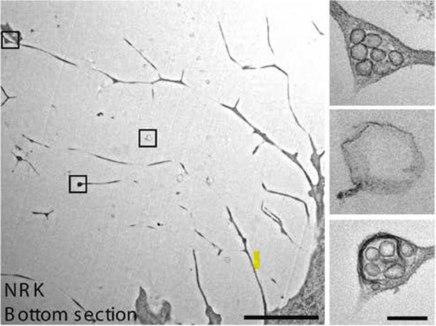
Package Left Behind. As rat kidney cells migrate across a cell culture plate (left), membrane bubbles (inset boxes) form on the retraction fibers they leave behind. Most of these blobs are stuffed with extracellular vesicles (right), giving them the look of a pomegranate interior. [Courtesy of Ma et al., Cell Research, 2015.]
Migrasomes were first identified a decade ago by scientists led by Li Yu at Tsinghua University-Peking University Joint Center for Life Sciences, Beijing. While studying rat kidney cells migrating across culture dishes, Yu and colleagues noticed bubble-like structures forming on the retraction fibers they left behind. These membrane spheres, which the authors dubbed migrasomes, ranged from 0.5 to 3 μm in diameter, and typically contained several vesicles, giving them a pomegranate-like appearance (image above). They were distinct from the multivesicular bodies that fuse with plasma membrane to release extracellular vesicles as, unlike those structures, they were devoid of the lysosomal marker LAMP1 (image below). Instead, migrasomes depended on the adhesion factor tetraspanin-4 (TSPAN4) for their formation (Ma et al., 2015).
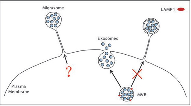
Distinct Release Pathways. Unlike the multivesicular bodies (MVB) that fuse with the plasma membrane to release exosomes, migrasomes do not contain the marker LAMP1 (red). Instead, they form on retraction fibers. [Courtesy of Ma et al., Cell Research, 2015.]
Scientists previously showed that mouse and human BAMs, too, produced migrasomes. Indeed, when Cai and colleagues exposed such cells to Aβ40, they formed retraction processes that contained migrasomes and AIM, aka apoptosis inhibitor of macrophage. They reported that this protein then attached to vascular amyloid and damaged brain blood vessels (Aug 2023 conference news; Hu et al., 2023).
In the new paper, joint first authors Mengyan Hu, Xinmei Kang, and Zhiruo Liu explored whether BAM migrasomes might influence brain aging in general. They found that BAMs in wild-type mice expressed senescence markers at around 6 months of age, well before other brain cell types became senescent. Senescent BAMs produced more than three times as many migrasomes as did those in younger mice, as measured using wheat germ agglutinin with a fluorescent tag. WGA binds to sialic acid and N-acetyl-D-glucosamine, which are enriched in migrasomes. In people, too, migrasome production by macrophages went up with age (image below).
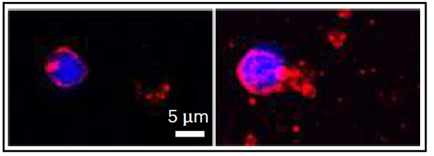
Old Macrophages Litter. In culture, monocyte-derived macrophages from a healthy 62-year-old (left) produce fewer migrasomes, measured with red fluorphore-tagged wheat germ agglutinin (red) than do those from an 80-year-old (right). Nuclei are blue. [Courtesy of Hu et al., Nature Aging, 2025.]
Hu and colleagues tied senescence, and its attendant migrasome explosion, to Aβ40. Starting when mice were 6 months old, they found, BAMs accumulated Aβ40, but not other forms of cellular waste, such as Aβ42 and α-synuclein. Exposing macrophages to Aβ40 in vitro made them senescent. Moreover, when Hu injected Aβ40 into the brains of 3-month-old mice, the peptide selectively accumulated in BAMs and almost doubled migrasome production.
What did these BAM migrasomes do? The authors isolated them from 18-month-old wild-type mice and injected them into 3-month-old ones. Lo and behold, their brain cells then became senescent, and the young mice developed memory problems. The authors traced this to AIM, the same factor that had damaged blood vessels in their earlier study. Migrasomes from macrophages lacking AIM no longer transmitted this senescence.
To figure out how AIM-filled migrasomes damaged the brain, the authors added a dye to the organelles and examined which brain cell types took them up. Microglia gobbled up most of them. About 10 percent of these microglia then became senescent, pro-inflammatory, and lost their phagocytic skills. AIM by itself produced the same effects in cultured microglia, showing that this protein was a likely culprit.
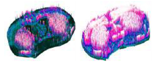
Fewer Migrasomes, More Wiring. In a coronal brain section from an 18-month-old mouse with its migrasomes suppressed (right), myelin (pink) was more abundant compared with an untreated peer (left). [Courtesy of Hu et al., Nature Aging, 2025.]
Finally, the authors prevented migrasome formation in aged mice by knocking down TSPAN4. Microglia remained youthful and phagocytic, and the mouse brains appeared healthier overall, maintaining more myelin (image above). They also seemed to function better. In memory tests, these mice did better than their untreated peers of the same age.
Whether migrasomes play a role in AD is unknown, though in 5xFAD mice, the authors found Aβ40 accumulating in BAMs and driving up migrasome production by 4 months of age, two months earlier than in wild-types. The data hint that amyloidosis could accelerate brain aging.
“Targeting perivascular macrophage-derived migrasomes could represent a novel therapeutic strategy to mitigate brain aging and the impact of amyloid pathology on the brain,” Iadecola suggested. Still, migrasomes may not be all bad. Bone marrow mesenchymal stem cells have been found to temper CAA in mice by forcing neutrophils to spew damaged mitochondria via migrasomes (Hu et al., 2025).—Madolyn Bowman Rogers
References
News Citations
Research Models Citations
Paper Citations
- Ma L, Li Y, Peng J, Wu D, Zhao X, Cui Y, Chen L, Yan X, Du Y, Yu L. Discovery of the migrasome, an organelle mediating release of cytoplasmic contents during cell migration. Cell Res. 2015 Jan;25(1):24-38. Epub 2014 Oct 24 PubMed.
- Hu M, Li T, Ma X, Liu S, Li C, Huang Z, Lin Y, Wu R, Wang S, Lu D, Lu T, Men X, Shen S, Huang H, Liu Y, Song K, Jian B, Jiang Y, Qiu W, Liu Q, Lu Z, Cai W. Macrophage lineage cells-derived migrasomes activate complement-dependent blood-brain barrier damage in cerebral amyloid angiopathy mouse model. Nat Commun. 2023 Jul 4;14(1):3945. PubMed. Correction.
- Hu M, Yi H, Wang S, Kang X, Liu Y, Liu Z, Huang H, Qin Q, Yuan L, Cai W, Qiu W, Lu Z, Liu S. Bone marrow mesenchymal stem cells protect against cerebral amyloid angiopathy by enhancing neutrophil mitocytosis. Neural Regen Res. 2025 Jun 19; Epub 2025 Jun 19 PubMed.
Further Reading
News
- Circulating Monocytes Replace Microglia, Border-Associated Macrophages
- Implicated in ARIA: Perivascular Macrophages and Microglia
- Does the Brain Use Microglia to Maintain Its Myelin?
- Macrophages Blamed for Vascular Trouble in ApoE4 Carriers
- When Perivascular Macrophages Spew SPP1, Microglia Eat Synapses
- Perivascular Macrophages: New Target in Aging and Alzheimer’s Disease?
- With Age, Macrophages Chew up the Blood-CSF Barrier
- Do Specialized Glycoproteins Prop Up Blood-Brain Barrier?
Primary Papers
- Hu M, Kang X, Liu Z, Wang S, Liu S, Li C, Lu D, Qin Q, Liu Y, Yi H, Yuan L, Liu Q, Lu Z, Cai W. Senescent-like border-associated macrophages regulate cognitive aging via migrasome-mediated induction of paracrine senescence in microglia. Nat Aging. 2025 Sep 10; Epub 2025 Sep 10 PubMed. Correction.
Annotate
To make an annotation you must Login or Register.
Comments
Weill College Medicine, New York
I find this paper very compelling. Our group and others have provided evidence that perivascular macrophages (PVMs) contribute to the detrimental effects of hypertension, Alzheimer’s disease pathology, cerebral amyloid angiopathy, stroke, and the ApoE4 genotype on the brain. However, the mechanisms by which PVMs influence the brain parenchyma have remained unclear. We have shown that PVMs generate substantial amounts of reactive oxygen species via NOX2 in response to Aβ, angiotensin, and ApoE4. The discovery here that PVMs in the context of aging and Aβ develop senescence and release migrasomes, which can propagate cellular senescence, offers a mechanistic link between PVMs senescence and parenchymal cells. This finding provides a potential explanation for how PVMs may exert broad effects on brain function.
Thus, targeting PVM-derived migrasomes could represent a novel therapeutic strategy to mitigate brain aging and the impact of amyloid pathology on the brain.
Mayo Clinic Florida
This study led by Wei Cai’s group proposes a new mechanism of aging, mediated by migrasomes, organelles of migratory cells, including border-associated macrophages (BAMs). BAMs have been reported to be constantly exposed to peripheral or brain Aβ, therefore expressing high levels of senescent genes in AD brains. Here, Hu et al. showed that BAM isolated from aged mice express significantly more TSPAN4, a migrasome marker, than BAMs isolated from young mice.
Notably, intracisternal injection of migrasomes isolated from aged mice caused cell cycle arrest, increased levels of senescence-associated beta-galactosidase, and a cognitive deficit as shown by the water maze test, in recipient mice as compared to mice injected with control beads or migrasomes isolated from young mice. Interestingly, the authors further showed that among all the cell types in the brain, microglia predominantly take up migrasomes, and develop senescent features upon receiving migrasomes isolated from Aβ-treated macrophages or aged mice.
These results advance the aging field by offering a clue to how aged cells may influence functions of other cell types via migrasomes. My only question is about the protocol to isolate migrasomes used in this article. It differs from protocols in previous papers, which used a sucrose gradient and ultra centrifugation approach (Zhang et al., 2023; Zhang et al., 2022). As migrasomes secrete exosomes when they are scissored, we need similar guidelines to the Minimal Information for Studies of Extracellular Vesicles created by the International Society of Extracellular Vesicles to enhance the rigor of studies using migrasomes (Welsh et al., 2024). Of note, the authors provide no structural assessment of the isolated migrasomes by electron microscopy, no quantification of the number of particles, nor evidence of enrichment or depletion of EV and non-EV markers, which would be helpful.
References:
Zhang X, Yao L, Meng Y, Li B, Yang Y, Gao F. Migrasome: a new functional extracellular vesicle. Cell Death Discov. 2023 Oct 18;9(1):381. PubMed.
Zhang C, Li T, Yin S, Gao M, He H, Li Y, Jiang D, Shi M, Wang J, Yu L. Monocytes deposit migrasomes to promote embryonic angiogenesis. Nat Cell Biol. 2022 Dec;24(12):1726-1738. Epub 2022 Nov 28 PubMed.
Welsh JA, Goberdhan DC, O'Driscoll L, Buzas EI, Blenkiron C, Bussolati B, Cai H, Di Vizio D, Driedonks TA, Erdbrügger U, Falcon-Perez JM, Fu QL, Hill AF, Lenassi M, Lim SK, Mahoney MG, Mohanty S, Möller A, Nieuwland R, Ochiya T, Sahoo S, Torrecilhas AC, Zheng L, Zijlstra A, Abuelreich S, Bagabas R, Bergese P, Bridges EM, Brucale M, Burger D, Carney RP, Cocucci E, Crescitelli R, Hanser E, Harris AL, Haughey NJ, Hendrix A, Ivanov AR, Jovanovic-Talisman T, Kruh-Garcia NA, Ku'ulei-Lyn Faustino V, Kyburz D, Lässer C, Lennon KM, Lötvall J, Maddox AL, Martens-Uzunova ES, Mizenko RR, Newman LA, Ridolfi A, Rohde E, Rojalin T, Rowland A, Saftics A, Sandau US, Saugstad JA, Shekari F, Swift S, Ter-Ovanesyan D, Tosar JP, Useckaite Z, Valle F, Varga Z, van der Pol E, van Herwijnen MJ, Wauben MH, Wehman AM, Williams S, Zendrini A, Zimmerman AJ, MISEV Consortium, Théry C, Witwer KW. Minimal information for studies of extracellular vesicles (MISEV2023): From basic to advanced approaches. J Extracell Vesicles. 2024 Feb;13(2):e12404. PubMed.
Make a Comment
To make a comment you must login or register.