
Do PERK’d Up Astrocytes Slow Amyloid Clearance?
Quick Links
Cells feature an array of biological cascades to respond to stressors such as protein aggregates, viruses, and DNA damage. When these pathways are activated too much or for too long, things can go awry. This may be the case for the unfolded protein response (UPR) in people with Alzheimer’s disease. In Neuron, May 21, scientists led by Guang Yang at Columbia University in New York and Zhongcong Xie at Massachusetts General Hospital reported that, in postmortem human brain and in mouse models of amyloidosis and tauopathy, the UPR—specifically the branch driven by protein kinase RNA-like ER kinase (PERK)—ramps up explicitly in astrocytes. This worsened pathology, in part by impeding glymphatic clearance, a waste-management system in the brain. Knocking out PERK in astrocytes, or blocking it, restored clearance, reduced amyloid pathology, and improved learning and memory, the authors report.
- Unfolded protein response mediator PERK is hyperactive in AD.
- The kinase impedes the brain’s glymphatic system.
- Could targeting the PERK-CK2-AQP4 axis swell glymphatic clearance, benefit cognition?
To the mind of Kaoru Yamada, University of Tokyo, the work supports the view that AD is not a neuron-centric disease but is largely driven by glial dysfunction. “This study reinforces the increasingly appreciated view that astrocytes play active and dynamic roles in maintaining brain homeostasis. It contributes meaningfully to our evolving understanding of glial function in AD,” Yamada wrote to Alzforum (comment below).

PERKed UP. Imaging and quantification of 5xFAD mouse brain (left) and postmortem human AD brain (right) reveals enhanced astrocytic inflammation seen by GFAP staining (green) and PERK phosphorylation (pink). [Image courtesy of Chen et al., Neuron, 2025.]
Glymphatic clearance, which relies on a network of astrocytic endfeet arranged along vessels of the cerebrovasculature, has been implicated in AD, though it is not without controversy. Perivascular fluid exchange throughout this network, primarily via the astrocytic channel aquaporin-4 (AQP4), has been reported to be essential for removal of both Aβ and tau (Aug 2012 news; Mar 2013 news; Feb 2022 news). Evidence from rodent and AD brain studies have shown that disrupting AQP4 expression, or its concentration at perivascular areas of the cell membrane, worsens both pathologies.
Although neuronal UPR signaling has been implicated in AD pathogenesis, the repercussions of its activation in astrocytes are unclear, note the authors. Given astrocytes’ essential role in supporting glymphatic clearance, they posited that the transcriptional changes brought on by excessive UPR activation may contribute to this system’s malfunction.

Clearing the Way. Time-lapse images of FITC-dextran tracer distribution in the mouse cortex show that while deleting PERK has no effect in wild-type (second row), it restores glymphatic flow in 5xFAD mice (bottom row). [Courtesy of Chen et al., Neuron, 2025.]
To test this, first author Kai Chen and colleagues knocked out astrocytic PERK in mice. While 5xFAD mice did not make much perivascular AQP4, PERK knockouts made more, almost as much as did wild-type controls. In vivo two-photon imaging of a cerebrospinal fluid tracer confirmed that while glymphatic flow in 5xFAD mice was weak, PERK deletion partially restored it. Moreover, deleting PERK from wild-type mice had no effect on AQP4 perivascular distribution or clearance of the tracer, suggesting that the kinase only impairs glymphatic clearance in the 5xFAD mice.
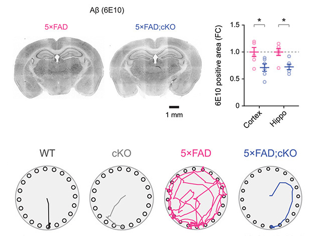
More PERK, More Problems. Knocking out PERK in astrocytes reduces amyloid plaque burden in 5xFAD (top). The knockouts more quickly found the escape hole in the Barnes maze test (bottom). [Courtesy of Chen et al., Neuron, 2025.]
Next, Chen and colleagues investigated what deleting PERK would do to Aβ, tau, inflammation, and cognition. The PERK-deleted 5xFAD mice had less plaque, less GFAP and IBA1—markers of activated astrocytes and microglia, respectively—and they better learned the location of an escape hole in the Barnes maze (image above). Blocking AQP4 with the selective inhibitor TGN-020 nullified these benefits, supporting the interpretation that they can be attributed to improved glymphatic function.
Additionally, the authors found that deleting PERK in PS19 transgenic mice, a model of tauopathy, restored dysfunctional glymphatic clearance, lowered phosphorylated and aggregated tau, and rescued cognitive deficits. Together, these findings suggest that repairing the PERK-CK2-AQP4 axis might help restore glymphatic flow in AD or other neurodegenerative diseases.
Glymphatic regulation is one of many astrocyte functions that likely influence AD pathologies, noted the authors. More work is needed to fully understand how astrocytic UPR and PERK signaling impacts AD progression. “While we identified CK2 as a potential mediator between PERK and AQP4, the mechanisms remain unclear, particularly how CK2 regulates AQP4 trafficking to astrocyte perivascular endfeet in AD,” Yang and Xie wrote to Alzforum (comment below). Using a protein-protein interaction model, they have identified potential CK2 phosphorylation sites on AQP4. They are now investigating which one is most important for AQP4 trafficking in the context of AD.—Anna Bright
Anna Bright is a Ph.D. student in New York City.
References
News Citations
- Brain Drain—'Glymphatic' Pathway Clears Aβ, Requires Water Channel
- Spinal Fluid Flush: Visualizing the Brain Drain With MRI
- Not Just Aβ—Glymphatic Flow Clears Tau, Too, Slowing Its Aggregation
Research Models Citations
Further Reading
News
- Not Just Aβ—Glymphatic Flow Clears Tau, Too, Slowing Its Aggregation
- Make Proteins, Save Memory: The Cellular Stress Response and Synapses
- Dearth of Water Channels a Sign of ‘Glymphatic’ Breakdown in Alzheimer’s?
- Brain Drain—'Glymphatic' Pathway Clears Aβ, Requires Water Channel
- PERKing Up Protein Synthesis May Prevent Neurodegeneration
Primary Papers
- Chen K, Morizawa YM, Nuriel T, Al-Dalahmah O, Xie Z, Yang G. Selective removal of astrocytic PERK protects against glymphatic impairment and decreases toxic aggregation of β-amyloid and tau. Neuron. 2025 Aug 6;113(15):2438-2454.e6. Epub 2025 May 21 PubMed.
Annotate
To make an annotation you must Login or Register.
Comments
The University of Tokyo
In this study, the authors clearly demonstrate that the ER stress/UPR mediator PERK plays a critical role in the altered glymphatic function associated with changes in AQP4 localization in AD model mice. By crossing PERK fl/fl, Aldh1l1-CreERT2 mice with either 5xFAD or PS19 models, they show not only a reduction in amyloid and tau pathology but also a rescue of associated behavioral alterations, which I found particularly compelling.
The mechanistic basis by which PERK disrupts glymphatic clearance appears to be complex. As the authors note in the discussion, additional factors such as neuroinflammation and the interplay between dural lymphatic drainage and glymphatic flow may also be involved. These findings highlight the multifactorial nature of clearance deficits in neurodegeneration.
This study reinforces the increasingly appreciated view that astrocytes play active and dynamic roles in maintaining brain homeostasis. It contributes meaningfully to our evolving understanding of glial function in AD.
The University of Texas Health Science Center at Houston
Columbia University
We were surprised to find that conditional knockout of PERK in astrocytes partially enhanced glymphatic function by restoring perivascular AQP4 localization. PERK activation is well-documented in Alzheimer’s disease, where it phosphorylates eIF2α and suppresses protein synthesis. While impaired protein synthesis in neurons and astrocytes has been reported in AD, the specific contribution of astrocytic protein synthesis to glymphatic function has been less explored. We were excited to find that PERK deletion in astrocytes improved glymphatic function in AD transgenic mice, suggesting that chronic activation of the PERK-eIF2α pathway may contribute to glymphatic failure in AD.
Our findings show that both genetic and pharmacological inhibition of PERK partially improved glymphatic function. This suggests that targeting the PERK signaling pathway could represent a novel therapeutic strategy to address glymphatic dysfunction, which is increasingly recognized in several neurodegenerative diseases, including AD, Parkinson’s disease, and amyotrophic lateral sclerosis.
Further investigation is planned. While we identified CK2 as a potential mediator between PERK and AQP4, the mechanisms remain unclear, particularly how CK2 regulates AQP4 trafficking to astrocyte perivascular endfeet in AD. Using a protein-protein interaction model, we have identified potential phosphorylation sites on AQP4 that CK2 may target. However, it remains to be determined which phosphorylation site is critical for AQP4 trafficking in the context of AD. In vitro cell culture systems may provide a more controlled environment to dissect the precise role of CK2 in modulating AQP4 dynamics.
Make a Comment
To make a comment you must login or register.