
Human Cortex Layers Up Sooner Than You Think
Quick Links
Perhaps our most defining characteristic as a species, the six-layered human cortex, hosts billions of neural connections that bestow Homo sapiens with higher-order thinking. But how does this “crowning achievement of evolution” take shape in the first place? With the help of spatial transcriptomics, scientists led by Christopher Walsh at Boston Children’s Hospital in Boston unveil the process at a new level of detail. Published May 14 in Nature, their study tracked the transcriptomes of individual neurons and other cells in situ within the emerging brain, focusing on the second and third trimesters of gestation. Using this technique, they observed a clear delineation of all cortical layers by five months, a full three months earlier than can been seen under a microscope. By 20 weeks of gestation, some regions of the brain, such as the primary and secondary visual cortices, had already partitioned into clearly defined subregions based on gene expression, yet still appeared like a homogenous sheet based on cellular architecture. The work suggests that cortical layers and regions start to form earlier than previously thought.
- Spatial transcriptomics charts development of the human brain.
- Cortical layers shape up three months before cellular architecture becomes visible.
- So does the delineation of primary and secondary visual cortices.
While focused on the developing brain, this study’s findings are relevant to understanding neurodegenerative diseases that arise decades later, commented Vivek Swarup of the University of California, Irvine. “This foundational atlas of cortical development, built using advanced spatial transcriptomic methods, is a valuable framework for identifying early developmental antecedents of disease susceptibility and resilience, informing future studies on AD mechanisms,” he wrote.
The dataset is available for sifting via an interactive browser at https://walshlab.org/research/cortexdevelopment/.
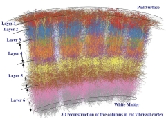
Behold, the Cortical Column. A three-dimensional rendering of the mammalian neocortex displays the six cortical layers—from the surface of the brain to the inside. Distinct cytoarchitectural features are visible via microscopy. [Underlying image from Marcel Oberländer, Beyond the Cortical Column, Neuroinformatics 2012, via Wikimedia Commons.]
The six layers of the mature neocortex—labeled 1 to 6 from surface to depth—are defined by distinctive populations of neurons and their axonal projections, such that each Neuroin can be clearly seen in cortical cross-sections via microscopy. For example, Layer 1, also known as the molecular layer, is outermost, and consists primarily of dendrites that project from neurons residing in deeper layers, along with sparse neuronal bodies. Layer 2, aka the external granular layer, is packed with small neurons called granule cells, while Layer 3, the external pyramidal layer, houses the cell bodies of pyramidal neurons. In turn, Layers 4 to 6—called the internal granular layer, the internal pyramidal layer, and the multiform or polymorphic layer, respectively—each have their own distinctive arrangements of neurons, projections, and connections. During development, these layers are thought to form from the inside out, such that the deeper layers form first, followed by the more superficial ones. Notably, none of these layers can be clearly delineated until near the end of human gestation.
To understand how neurons migrate into these various layers, co-first authors Xuyu Qian, Kyle Coleman, Shunzhou Jiang, and colleagues sampled cross-sections of eight major cortical areas from 10 fetal brains, obtained from the Department of Pathology at Beth Israel Deaconess Medical Center in Boston or the University of Maryland Brain and Tissue Bank. The fetuses ranged from gestational weeks 15 to 34. Six brains were included in an initial analysis, four more added later. The scientists used MERFISH, a microscopy-based spatial transcriptomics technique that uses fluorescent nucleic acid probes to image the abundance of some 300 transcripts at near-single-cell resolution. Because fetal brain tissue is so dense, the scientists designed and ran a deep-learning model to improve precision.
Among the 16 million cell transcriptomes that were spatially mapped across the samples, the scientists identified eight classes of cells, which could be broken down into 33 transcriptional cell types. These, in turn, could be split into 114 subtypes, including 58 different excitatory neurons.
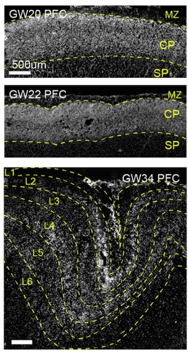
Latent Layers. At gestational weeks 20 (top) and 22 (middle), nuclear staining visualizes only the marginal zone (MZ), cortical plate (CP), and subplate (SP). By week 34 (bottom), the marginal zone and cortical plate have morphed into the six cortical layers. [Courtesy of Qian et al., Nature, 2025.]
In addition to MERFISH, the scientists also used traditional nuclei staining to capture the overall cytoarchitecture of cells in the brain. Consistent with previous reports, at 20 and 22 weeks old, they were able to discern only three morphological layers: the marginal zone, cortical plate, and subplate (image at right). During development, neurons from the marginal zone go on to form Layer 1, and other neurons from the subplate and cortical plate move into position to form the other five cortical layers. It was not until week 34 that the six cortical layers became visible via nuclear staining.
However, when spied through the lens of spatial transcriptomics, nascent cortical layers started taking shape as early as week 15. The scientists observed these budding layers by mapping the locations of the 58 excitatory neuron subtypes, some of which ended up in one specific cortical layer by week 34. Strikingly, at week 15, some of these neurons had already started taking up positions in the cortical plate corresponding to their final location within the cortex. This migration was further refined at weeks 20 and 22, at which point all six layers could clearly be seen within the cortical plate (image below). Consistent with the inside-out development of the cortex, the inner layers started shaping up before the outer ones.
To Walsh, the findings show that major features of the fate, or identity, of brain cells are fixed when they are first formed, and are then fine-tuned for months after.
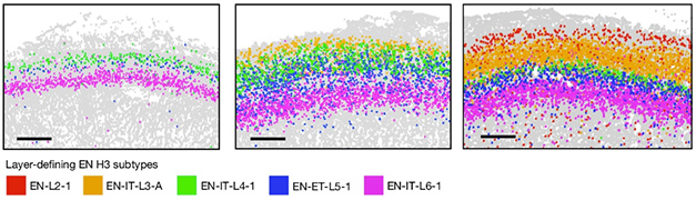
Hello, Layers! In this cross-section of prefrontal cortex, layer-defining excitatory neurons (EN) start to move into position within the cortical plate by gestational week 15 (left), when Layers 4 (green) and 5 (blue) separate from Layer 6 (pink). Layer 3 (orange) emerges at week 20 (middle), while Layer 2 (red) forms at week 22. Layer 1, which forms earlier from the marginal zone, is not shown. [Courtesy of Qian et al., Nature, 2025.]
During the third trimester, neurons are thought to develop their “personalities.” Consistent with this, the number of distinct subtypes of excitatory neuron increased dramatically between weeks 22 and 34. For example, one cluster of Layer-6-defining excitatory neurons seen at week 22 had branched out into nine clusters by week 34.
Besides timing the layering of the cortex, spatial transcriptomics also uncovered early distinctions between individual brain regions. A stunning example? Demarcation of the V1 and V2 regions of the visual cortex. Located within the occipital cortex at the back of the brain, and spanning all six cortical layers, the mature visual cortex includes five subregions, V1 to V5, which receive and process visual information transmitted from the optic nerve. Signals pass from the retinas to the thalamus to the lateral geniculate nucleus before synapsing with neurons in V1. Also known as the primary visual cortex, V1 responds to simple visual information such as orientation and direction. It passes on these signals to V2, which hands them off to V3, and so on. Each subsequent region encodes and integrates increasingly complex visual components. At week 20, there were no obvious anatomical differences between V1 and V2 à la light microscopy. However, spatial transcriptomics revealed an abrupt boundary between them, based on eight subtypes of excitatory neurons (image below).
This V1-V2 schism was most pronounced in Layers 3 and 4. Notably, these layers receive the inputs from the retina by way of the thalamus and lateral geniculate nucleus. The scientists proposed that the completion of this optic circuit might spark a transcriptional switch in V1 and V2 neurons, which facilitates the distinct functions of the two regions. They noted that this switch precedes the moment when the developing fetus first opens its eyes, at around week 26.

Hidden Borders. At week 20, regions V1 and V2 of the visual cortex exhibit no morphological differences (top), but spatial transcriptomics clearly delineated these regions by distinct neuronal subtypes, especially in Layers 3 and 4. [Courtesy of Qian et al., Nature, 2025.]
The scientists went on to identify specific genes involved in different stages of brain development, integrating their spatial transcriptomics data with single-cell RNA sequencing to study the entire transcriptome. Those findings are available for perusal online.
What might all of this portend, if anything, about age-related neurodegenerative diseases decades later? Walsh told Alzforum that although there are no obvious examples in this study, it’s clear that development and degeneration are connected. For example, his group has demonstrated that somatic mutations that arise in ALS/FTD genes during development might beckon those diseases later in life (Zhou et al., 2023). Other scientists have reported that processes that are part and parcel of normal brain development, such as microglial pruning of synapses, can reawaken and go haywire in the context of neurodegenerative disease (Stevens et al., 2007; Paolicelli et al., 2011; Mar 2015 conference news).
Swarup suggested that the way region-specific synaptic circuitry gets wired up might predispose some regions to disease while conferring resilience to others (Aug 2015 conference news; Apr 2017 news). “Moreover, disruptions or subtle deviations in these developmental trajectories might underpin regional susceptibilities characteristic of AD and other neurodegenerative disorders,” he wrote.—Jessica Shugart
References
News Citations
- Microglia Rely on Mixed Messages to Select Synapses for Destruction
- Does Brain Development in Childhood Set the Stage for Dementia?
- Childhood Epilepsy Begets Adult Amyloid
Paper Citations
- Zhou Z, Kim J, Huang AY, Nolan M, Park J, Doan R, Shin T, Miller MB, Chhouk B, Morillo K, Yeh RC, Kenny C, Neil JE, Lee C-Z, Ohkubo T, Ravits J, Ansorge O, Ostrow LW, Lagier-Tourenne C, Lee EA, Walsh CA. Somatic Mosaicism in Amyotrophic Lateral Sclerosis and Frontotemporal Dementia Reveals Widespread Degeneration from Focal Mutations. 2023 Dec 01 10.1101/2023.11.30.569436 (version 1) bioRxiv.
- Stevens B, Allen NJ, Vazquez LE, Howell GR, Christopherson KS, Nouri N, Micheva KD, Mehalow AK, Huberman AD, Stafford B, Sher A, Litke AM, Lambris JD, Smith SJ, John SW, Barres BA. The classical complement cascade mediates CNS synapse elimination. Cell. 2007 Dec 14;131(6):1164-78. PubMed.
- Paolicelli RC, Bolasco G, Pagani F, Maggi L, Scianni M, Panzanelli P, Giustetto M, Ferreira TA, Guiducci E, Dumas L, Ragozzino D, Gross CT. Synaptic pruning by microglia is necessary for normal brain development. Science. 2011 Sep 9;333(6048):1456-8. PubMed.
External Citations
Further Reading
No Available Further Reading
Primary Papers
- Qian X, Coleman K, Jiang S, Kriz AJ, Marciano JH, Luo C, Cai C, Manam MD, Caglayan E, Lai A, Exposito-Alonso D, Otani A, Ghosh U, Shao DD, Andersen RE, Neil JE, Johnson R, LeFevre A, Hecht JL, Micali N, Sestan N, Rakic P, Miller MB, Sun L, Stringer C, Li M, Walsh CA. Spatial transcriptomics reveals human cortical layer and area specification. Nature. 2025 May 14; Epub 2025 May 14 PubMed.
Annotate
To make an annotation you must Login or Register.
Comments
Rush University
This is a fascinating study, encompassing over 18 million single cells across eight cortical areas and seven developmental time points. By combining MERFISH with a deep-learning-based nuclear segmentation pipeline, the authors created an extensively detailed, spatially resolved single-cell atlas of the developing human cerebral cortex.
The study provides compelling evidence for a continuous anterior-posterior gradient of molecular and cellular features, supporting the classical protomap model of cortical arealization. Remarkably, it also identifies a sharp molecular boundary between the primary visual cortex (V1) and secondary visual cortex (V2) as early as gestational week 20 in post-migratory excitatory neurons. This finding supports aspects of the protocortex hypothesis, suggesting that while many cortical areas emerge through gradual transitions, specific boundaries, such as between V1 and V2, can be sharply defined early in development.
Beyond developmental insights, this study also showcases the potential of cross-modal gene expression imputation, such as the ENVI method, to align spatial and single-cell transcriptomic data. While this approach has not yet been fully leveraged in disease models, it holds great promise for application to postmortem human brains. Extending this strategy to neurodegenerative or neurodevelopmental disease contexts could uncover how spatial patterning and cell-type composition are disrupted during pathological progression, offering a powerful framework for future studies.
Make a Comment
To make a comment you must login or register.