
Introducing Next-Gen AAVs, Now Equipped with Enhancers
Quick Links
A new class of AAVs has arrived. In six coordinated studies published on 21 May across Neuron, Cell, Cell Reports, and Cell Reports Methods, scientists unveil viral vectors fitted with cell-type-specific enhancers that deliver gene expression with unprecedented precision. Developed as part of the National Institutes of Health’s Brain Research Through Advancing Innovative Neurotechnologies (BRAIN) Initiative, this suite of viruses is available for immediate use.
- Cell-specific enhancers improve specificity of AAVs.
- Six studies demo these latest vectors.
- Constructs are available through Addgene and the Allen Institute.
Adeno-associated viral (AAV) vectors have become indispensable tools for labeling, tracing, and manipulating cells. These molecular workhorses can deliver everything from fluorescent reporters and calcium indicators to recombinases, optogenetic switches, and therapeutic genes. However, AAV-based therapies have seen mixed results in clinical trials, prompting some companies to abandon AAV research altogether. A key challenge lies in poor tissue targeting and lack of expression control, issues that have contributed to clinical trial setbacks and safety concerns (Duan, 2023). Most AAVs rely on broadly active promoters, which often drive transgene expression in unintended cell types.
To tackle this problem, a consortium of scientists turned to enhancers—short, noncoding DNA sequences that serve as regulatory elements. When bundled into an AAV construct, enhancer elements will activate gene expression only in cells whose transcriptional machinery recognize and respond to them. This strategy is showcased in this series of studies, which are available on Cell’s BRAIN Armamentarium landing page and reviewed by Bosilijka Tasic and Gord Fishell in Neuron, May 21. Each study targets a distinct cell type within the nervous system.
Four of the papers are published in Neuron. In one, scientists led by Xiangmin Xu at the University of California, Irvine, scoured CATlas, a repository of regulatory elements derived from single-cell epigenomic datasets, to track down enhancers in brain endothelial cells. They packaged these into AAV vectors and tested them in several different mouse strains, including the 5xFAD model of amyloidosis.The scientists injected the vectors into blood vessels behind the eyes—a method that enables rapid delivery to the brain. They examined the brains of 1-year-old 5xFAD mice, four weeks after injection. In plaque-dense regions, the vectors robustly labelled endothelial cells with minimal off-target expression in other cell types (Velazquez-Rivera et al., 2025) (image below).
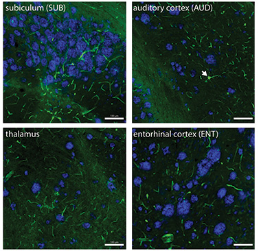
Endothelial Cells Shine Amidst Plaques. Endothelial cells targeted by AAV glow green, while Aβ plaques appear blue across various brain regions of 5xFAD mice. The white arrowhead points to an occasional stray labeled neuron. [Courtesy of Velazquez-Rivera et al., Neuron, 2025.]
In another Neuron study, scientists led by Yating Wang, Gord Fishell, and Jordane Dimidschstein at the Broad Institute in Cambridge, Massachusetts, set their sights on interneurons. They combed through single-cell datasets from mouse brain tissue to identify enhancer sequences located in regions of open chromatin. By packaging these enhancer sequences into AAVs, the team was able to drive expression in distinct cortical and striatal interneuron populations.
The research groups of William Stauffer and Andreas Pfenning at the University of Pittsburgh were also on the hunt for cortical enhancers. After mining single-cell epigenomic data from rhesus macaques to identify open chromatin regions, they used machine learning to whittle these down to the most promising cell-specific enhancers. Also reported in Neuron, their top picks successfully restricted channelrhodopsin expression to pyramidal neurons in Layers 2 and 3 of the monkey cortex.
Bosiljka Tasic and colleagues at the Allen Institute, Seattle, also identified enhancer sequences that drive expression in cortical cells. These scientists used single-nuclei transcriptomic and epigenomic datasets from mouse and human samples to uncover regulatory elements active in in the cortex. This effort yielded more than 1,000 different enhancer AAV vectors covering most major cortical neurons.
Jonathan Ting and colleagues, also at the Allen Institute, focused deeper in the brain and set their sights on the striatum, a region notoriously difficult to target with cell-type specificity due to its tightly interwoven mix of neurons. Undeterred, the team identified and validated enhancers that selectively drive expression in either D1- or D2-expressing spiny neurons, as well as all major classes of interneurons. The vectors performed reliably in both mice and monkeys.
Tanya Daigle’s team identified enhancers that enabled precise targeting of distinct subpopulations within spinal motor neuron pools. As described in Cell Reports, their top-performing AAVs drove fluorescent reporter expression in spinal motor neurons across mice, rats, and monkeys.
Finally, Xu and colleagues took another look at a capsid once thought to specifically target microglia. They discovered that it actually lights up excitatory neurons. When expressing a fluorescent reporter, the vector turned out to be an effective tool for mapping neural circuits in the mouse hippocampus. Their study rounded out the series and was published in Cell Reports Methods.
Each study is accompanied by standard operating procedures and user guides for deploying these newfangled AAVs. The full toolkit is available through Addgene and the Allen Institute’s Genetic Tools Atlas.—George R. Heaton
George Heaton is a freelance writer in Durham, North Carolina.
References
Research Models Citations
Paper Citations
- Duan D. Lethal immunotoxicity in high-dose systemic AAV therapy. Mol Ther. 2023 Nov 1;31(11):3123-3126. Epub 2023 Oct 10 PubMed.
- Tasic B, Fishell G. Exploring brain circuits, one cell type-or more- at a time. Neuron. 2025 May 21;113(10):1469-1473. PubMed.
- Velazquez-Rivera E, Dey O, Kim NS, Cao W, Ye Q, Gao P, Thai A, Nguyen JK, Zhang H, Ting JT, Gopi M, Ren B, Holmes TC, Xu X. Specific targeting of brain endothelial cells using enhancer AAV vectors. Neuron. 2025 May 21;113(10):1562-1578.e6. PubMed.
External Citations
Further Reading
No Available Further Reading
Primary Papers
- Tasic B, Fishell G. Exploring brain circuits, one cell type-or more- at a time. Neuron. 2025 May 21;113(10):1469-1473. PubMed.
- Furlanis E, Dai M, Leyva Garcia B, Tran T, Vergara J, Pereira A, Gorissen BL, Wills S, Vlachos A, Hairston A, Dwivedi D, Du S, McMahon J, Huang S, Morabito A, Vazquez A, Kim S, Lee AT, Chang EF, Razzaq T, Qazi A, Vargish G, Yuan X, Caccavano A, Hunt S, Chittajallu R, McLean N, Hewitt L, Paranzino E, Rice H, Cummins AC, Plotnikova A, Mohanty A, Tangen AC, Shin JH, Azadi R, Eldridge MA, Alvarez VA, Averbeck BB, Alyahyay M, Vallejo TR, Soheib M, Vattino LG, MacGregor CP, Chatain CP, Banks E, Olah VJ, Naskar S, Hill S, Liebergall S, Badiani R, Hyde L, Hanley E, Xu Q, Allaway KC, Goldberg EM, Rowan MJ, Nowakowski TJ, Lee S, Favuzzi E, Kaeser PS, Sjulson L, Batista-Brito R, Takesian AE, Ibrahim LA, Iqbal A, Pelkey KA, McBain CJ, Dimidschstein J, Fishell G, Wang Y. An enhancer-AAV toolbox to target and manipulate distinct interneuron subtypes. Neuron. 2025 May 21;113(10):1525-1547.e15. PubMed.
- He J, Phan BN, Kerkhoff WG, Alikaya A, Hong T, Brull OR, Fredericks JM, Sedorovitz M, Srinivasan C, Leone MJ, Wirfel OM, Brown A, Dauby S, Tittle RK, Lin MK, Hooks BM, Bostan AC, Gharbawie OA, Byrne LC, Pfenning AR, Stauffer WR. Machine learning identification of enhancers in the rhesus macaque genome. Neuron. 2025 May 21;113(10):1548-1561.e8. PubMed.
- Hunker AC, Wirthlin ME, Gill G, Johansen NJ, Hooper M, Omstead V, Vargas S, Lerma MN, Taskin N, Weed N, Laird WD, Bishaw YM, Bendrick JL, Gore BB, Ben-Simon Y, Opitz-Araya X, Martinez RA, Way SW, Thyagarajan B, Otto S, Sanchez RE, Alexander JR, Amaya A, Amster A, Arbuckle J, Ayala A, Baker PM, Barcelli T, Barta S, Bertagnolli D, Bielstein C, Bishwakarma P, Bowlus J, Boyer G, Brouner K, Casian B, Casper T, Chakka AB, Chakrabarty R, Chong P, Clark M, Colbert K, Daniel S, Dawe T, Departee M, DiValentin P, Donadio NP, Dotson NI, Dwivedi D, Egdorf T, Fliss T, Gary A, Goldy J, Grasso C, Groce EL, Gudsnuk K, Han W, Haradon Z, Hastings S, Helback O, Ho WV, Huang C, Johnson T, Jones DL, Juneau Z, Kenney J, Leibly M, Li S, Liang E, Loeffler H, Lusk NA, Madigan Z, Malloy J, Malone J, McCue R, Melchor J, Mich JK, Moosman S, Morin E, Naidoo R, Newman D, Ngo K, Nguyen K, Oster AL, Ouellette B, Oyama AA, Pena N, Pham T, Phillips E, Pom C, Potekhina L, Ransford S, Ray PL, Reding M, Rette DF, Reynoldson C, Rimorin C, Sigler AR, Rocha DB, Ronellenfitch K, Ruiz A, Sawyer L, Sevigny JP, Shapovalova NV, Shepard N, Shulga L, Soliman S, Staats B, Taormina MJ, Tieu M, Wang Y, Wilkes J, Wood T, Zhou T, Williford A, Dee N, Mollenkopf T, Ng L, Esposito L, Kalmbach BE, Yao S, Ariza J, Collman F, Mufti S, Smith K, Waters J, Ersing I, Patrick M, Zeng H, Lein ES, Kojima Y, Horwitz G, Owen SF, Levi BP, Daigle TL, Tasic B, Bakken TE, Ting JT. Enhancer AAV toolbox for accessing and perturbing striatal cell types and circuits. Neuron. 2025 May 21;113(10):1507-1524.e17. PubMed.
- Kussick E, Johansen N, Taskin N, Chowdhury A, Quinlan MA, Fraser A, Clark AG, Wynalda B, Martinez R, Groce EL, Reding M, Liang E, Shulga L, Huang C, Casper T, Clark M, Ho W, Gao Y, van Velthoven CT, Sobieski C, Ferrer R, Berg MR, Curtis BC, English C, Day JC, Fortuna MG, Donadio N, Newman D, Yao S, Chakka AB, Goldy J, Torkelson A, Guzman JB, Chakrabarty R, Nguy B, Guilford N, Pham TH, Wright V, Ronellenfitch K, Naidoo R, Kenney J, Williford A, Ramakrishnan C, Drinnenberg A, Gudsnuk K, Thyagarajan B, Smith KA, Dee N, Deisseroth K, Zeng H, Yao Z, Tasic B, Levi BP, Hodge R, Bakken TE, Lein ES, Ting JT, Daigle TL. Enhancer AAVs for targeting spinal motor neurons and descending motor pathways in rodents and macaque. Cell Rep. 2025 May 20;:115730. Epub 2025 May 20 PubMed.
- Cao W, Tan Z, Berackey BT, Nguyen JK, Brown SR, Du S, Lin B, Ye Q, Seiler M, Holmes TC, Xu X. An AAV capsid proposed as microglia-targeting directs genetic expression in forebrain excitatory neurons. Cell Rep Methods. 2025 May 13;:101054. Epub 2025 May 13 PubMed.
Annotate
To make an annotation you must Login or Register.
Comments
REGENXBIO
These reports describe recombinant AAVs that express reporter genes under the control of cell-specific transcriptional enhancers (enhancer rAAV), and their use to label target cells in the CNS with a high level of specificity and sensitivity. The approach is validated in cortical, striatal, and spinal cord neurons, as well as in non-neuronal cells of the glia and the vascular endothelium.
Somatic AAV gene transfer of reporter or therapeutic transgenes has numerous advantages over the current gold-standard methods using transgenic animals with Cre-mediated induction of cell specific reporters. Above all, enhancer rAAVs can be used to probe and manipulate brain circuits in different species, including primates, whereas the transgenic studies are mostly confined to rodents.
This new tool kit is exciting because it will enable studies of brain connectivity and development in ways that have not been possible up to now. It will also help design more precise and safer therapeutic modalities in the CNS. The new AAV-based tools are validated by measuring, in the target cells of the same animal, the signal generated by chromosomal Cre-mediated expression, and the one obtained from the rAAV genome. A high level of specificity and sensitivity is generally reported. However, these numbers vary, depending on the target cell type and the enhancer rAAV used. Specificities as low as 20 percent are reported, pointing to limitations of the approach.
In discussing these shortcomings, the authors note that the outcome of the studies can vary with the dose of rAAV, the age of the animal, and the route of administration. Also, the introduction of multiple copies of an enhancer sequence in a cell can induce squelching of transcription factors. Although no evidence of this is reported, it is indeed a source of concern, and it may be enhancer dependent.
Users of the technology should keep in mind that other factors are likely to limit or bias the precision of the rAAV tools. Toxicities associated with the quality of the rAAV preparation can have an impact, calling for careful analysis of vector quality. Certain cell types may not be transduced efficiently (or at all) when using certain capsids. Furthermore, it is now well-demonstrated that capsid proteins themselves can radically influence the formation of chromatin around the rAAV genome when it is delivered to the nucleus of the target cell. Cases of complete transcriptional shut-off have been described and that may counteract the activity of the enhancers. This bias can be cell- and species-specific.
With such a powerful tool kit, future investigators will need to go through a detailed validation for each novel combination of rAAV, enhancer, species, and route of administration. The technology is groundbreaking but not (yet) turnkey.
Make a Comment
To make a comment you must login or register.