
Multiscale Proteomics of Parahippocampal Gyrus Uncovers Drivers of AD
Quick Links
In a new proteomics extravaganza, scientists have unearthed groups of proteins that appear to be at the heart of Alzheimer’s disease pathogenesis. As described in Cell on September 25, scientists led by Bin Zhang and Dongming Cai of the Icahn School of Medicine at Mount Sinai in New York, and Junmin Peng of St. Jude Children’s Hospital in Memphis, Tennessee, deployed deep proteomic profiling of parahippocampal gyri from nearly 200 people who had died at various stages of AD. This region is impacted early, and severely, in the progression of the disease. By corroborating their proteomics data with complementary genomic and transcriptomic data from a broader set of brain samples, the scientists uncovered networks of co-expressed proteins that associated both with AD pathology and with cognitive symptoms.
- Proteomic, transcriptomic, and genetic data from hundreds of brain samples tie protein networks to AD.
- A clique of co-expressed proteins reflects toxic cross talk among astrocytes, microglia, and neurons.
- AHNAK, an astrocytic scaffold protein, drives this network.
One protein clique appeared to reflect an ongoing neurotoxic conversation among different cell types, including neurons, microglia, and astrocytes. AHNAK—a scaffolding protein expressed by astrocytes—emerged as an orchestrator of this cellular symphony. Knocking down this troublemaker in astrocytes spared co-cultured neurons, lowered phosphorylated tau, and turned down other seemingly detrimental inflammatory and metabolic pathways. Besides AHNAK, the study uncovered hundreds of other “key driver proteins” at work in other cell-type-specific networks, which the authors view as potential therapeutic targets.
Because this study tied proteins, rather than transcripts, to disease, the findings are more directly applicable to drug discovery, Zhang told Alzforum. “We encourage the scientific community to investigate these driver proteins—because one lab can only do so much,” he said.
Proteomic technology has steadily advanced in recent years, allowing scientists to take stock of ever more proteins and survey more regions of the brain (May 2020 news; Feb 2022 news). However, most proteomic analyses have centered on regions, such as the prefrontal cortex, that become inundated with AD pathology only in the later stages of disease. Until now, no large-scale, in-depth proteomics analysis had been done on the parahippocampal gyrus (PHG), a region affected early in AD (Wang et al., 2016).
To tackle this, co-first authors Erming Wang, Kaiwen Yu, and Jiqing Cao surveyed the proteomes of PHG tissue from 185 people in the Mount Sinai Brain Bank cohort. Based on the CERAD classification of AD neuropathology, these samples came from 60 non-diseased controls, 24 with possible AD, 25 with probable AD, and 76 with definite AD. Liquid chromatography-tandem mass spectrometry detected 12,147 unique protein isoforms, expressed by 9,272 genes, across all 185 samples. They then investigated how levels of each protein related to the severity of different neuropathological and clinical measures, including Aβ plaque burden, CERAD score, Braak stage, and clinical dementia rating (CDR) scale. For each of these traits, they identified at least 1,000 differentially abundant proteins that tracked with severity. On average, proteins that increased with severity tended to be involved in glial and/or immune pathways, while proteins that were turned down as disease worsened take part in neuronal processes such as synaptic signaling.
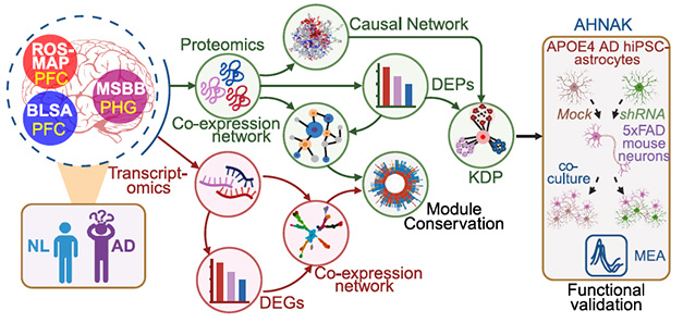
Omics Intersection. Proteomic and transcriptomic data from three brain sample cohorts (left) identified networks of co-expressed proteins that correlated with features of AD (middle). Causal analyses identified key driver proteins (KDPs), including AHNAK, that were validated in cultured astrocytes (right). [Courtesy of Wang et al., Cell, 2025.]
Around 60 percent of the proteins associated with AD neuropathology, as gauged by CERAD, overlapped with those that tracked with dementia severity, suggesting both common and distinct pathways involved in pathology and its cognitive consequences. The suite of AD-related proteins also differed substantially by sex, with both overlapping and sex-specific signatures.
The scientists also looked at how APOE genotype modulated parahippocampal protein profiles. They compared the proteomes of ApoE3 carriers with or without AD, with proteomes of ApoE4 carriers with AD. They did not have enough samples to look at ApoE4 carriers without AD. In a nutshell, they found 42 differentially expressed proteins when comparing ApoE3 carriers with AD to those without AD, while more than 1,300 proteins differed between ApoE4 carriers with AD and ApoE3 carriers without AD. Strikingly, nary a single protein differed substantially between ApoE3 and ApoE4 carriers with AD. This aligns with the idea that ApoE4 hastens onset of AD, but once the disease process is set in motion, brain changes are similar regardless of APOE genotype, Zhang said.
For the most part, the disease-related proteomic profiles Wang and colleagues identified in the PHG significantly overlapped with those reported in prefrontal cortex (PFC) samples surveyed in two other cohorts: ROSMAP and the Baltimore Longitudinal Study of Aging (BLSA). Wang also cross-referenced their proteomic findings with transcriptomic data. Within the PHG dataset, just under half of upregulated proteins and nearly 80 percent of downregulated proteins tracked with concomitant changes at the mRNA level.
To get a handle on how these complex proteomic changes relate to biological function, the scientists used statistical algorithms to group all the proteins detected in their analyses into bins of co-expressed proteins. This produced a windfall of 386 modules, each including 10 to 3,118 proteins. To suss out which of these modules were most relevant for AD, the scientists assessed how stocked each was with differentially expressed, AD-associated proteins. They were then able to rank the co-expression modules based on the strength of their ties to AD.
Among the top 30 AD-linked modules, many comprised proteins predominantly expressed by one cell type or another. While glia-specific modules tended to be upregulated, neuron-specific versions tended to be turned down. Strikingly, the top AD hit module—M3—included neuronal proteins that were turned down, as well as astrocytic and microglial proteins that were cranked up. These 3,118 co-regulated proteins were involved in a variety of functions, including metabolism, antigen presentation, and vesicular trafficking. When put in the context of the other top AD-associated modules, M3 stood out as a common thread connecting them all (image below).
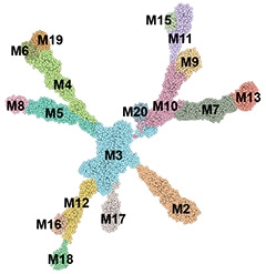
All Roads Lead to M3. Among the top modules of co-expressed proteins that tracked with AD, M3 was most strongly associated with disease and most connected to proteins in the other modules. [Courtesy of Wang et al., Cell, 2025.]
Zhang was struck by this result. “It implies that M3 is so central; it dominates disease-related changes across the proteome,” he said. He added that this was the first time such a multicellular module had been ranked above all others, including single-cell modules, in its relationship to AD.
M3 proteins also associated with AD in PFC samples from the ROSMAP cohort, but not as strong as in the PHG. This could be because the PHG is affected earlier in the disease process, and more severely, Zhang said.
Some of the other top AD-associated modules also contained thousands of proteins. To zero in on which ones dictate co-expression, the scientists integrated multiple layers of proteomic and genomic data to infer causal relationships among them. They included transcriptomic data, transcription factor-target relationships, and expression quantitative trait loci. This computational wizardry culminated in a “key driver analysis,” which identified 580 key driver proteins (KDPs), or top regulatory positions, in these disease networks.
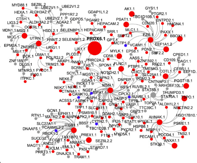
Network Masters. Multi omic analysis inferred causal relationships among proteins within networks controlled by key driver proteins (large circles). A subnetwork of up- (red) and downregulated (blue) proteins controlled by the two most powerful drivers—PRDX6.1 and MSN.1—is shown above. AHNAK (bottom right) is part of this subnet. [Courtesy of Wang et al., Cell, 2025.]
Some of these head honchos, such as PRDX6 and moesin (MSN), had been implicated in AD or neurodegeneration before. For example, PRDX6, the top-ranking KDP, reportedly hampers neurogenesis in the context of neurodegeneration, and exerts anti-inflammatory effects (Yeo et al., 2019). MSN is part of the ezrin, radixin, moesin complex that tethers the plasma membrane to the actin cytoskeleton, and reportedly plays a part in glial metabolism (Vega et al., 2018; Johnson et al., 2020).
However, the vast majority of the KDPs had been minimally studied or not investigated in the context of AD. One these—AHNAK—drove the central M3 network. Single-nucleus RNA sequencing data from the PHG samples indicated that it is primarily expressed by astrocytes.
To learn how AHNAK might orchestrate disease-related neuron-glia crosstalk, the scientists used shRNA to knock it down in iPSC-derived human astrocytes. Because they found that AHNAK expression was highest in ApoE4 carriers with AD, they generated these astrocytes from an ApoE4/E4 carrier. In a nutshell, astrocytes deprived of AHNAK had about 25 percent less phosphorylated tau, and, when co-cultured with neurons from 5xFAD mice, these AHNAK-less astrocytes revitalized sluggish synaptic firing observed in these neurons. What’s more, the scientists found that knocking down AHNAK reversed many disease-associated proteomic changes in astrocytes, restoring levels of proteins involved in metabolism, MAPK signaling, inflammation, and lipid metabolism.
The findings paint AHNAK as the ringleader in AD-associated proteomic changes, but the mechanisms need further study, Zhang told Alzforum. Also known as desmoyokin, AHNAK, a massive scaffolding protein, interacts with numerous partners. Considered a jack of all trades, it has been implicated in tumor suppression, modulation of calcium signaling, immune regulation, and development of the blood brain barrier (Davis et al., 2014; Zhang et al., 2023; Sundararaj et al., 2021). More than a decade ago, a proteomic analysis identified AHNAK among the top upregulated proteins in AD brain samples (Manavalan et al., 2013). Later, it was implicated as a ringleader in AD-associated gene-expression networks active in the human hippocampus, and, in mice, as a mediator of stress resilience in the prefrontal cortex (Chen et al., 2024).
Besides AHNAK, numerous other driver proteins are ripe for investigation and could yield promising therapeutic targets, Zhang said.—Jessica Shugart
References
News Citations
- Massive Proteomics Studies Peg Glial Metabolism, Myelination, to AD
- Proteomics Highlight Alzheimer’s Changes in Matrisome, MAPK Signaling
Paper Citations
- Wang M, Roussos P, McKenzie A, Zhou X, Kajiwara Y, Brennand KJ, De Luca GC, Crary JF, Casaccia P, Buxbaum JD, Ehrlich M, Gandy S, Goate A, Katsel P, Schadt E, Haroutunian V, Zhang B. Integrative network analysis of nineteen brain regions identifies molecular signatures and networks underlying selective regional vulnerability to Alzheimer's disease. Genome Med. 2016 Nov 1;8(1):104. PubMed.
- Yeo IJ, Park MH, Son DJ, Kim JY, Nam KT, Hyun BK, Kim SY, Jung MH, Song MJ, Chun HO, Lee TH, Han SB, Hong JT. PRDX6 Inhibits Neurogenesis through Downregulation of WDFY1-Mediated TLR4 Signal. Mol Neurobiol. 2019 May;56(5):3132-3144. Epub 2018 Aug 10 PubMed.
- Vega IE, Umstead A, Wygant CM, Beck JS, Counts SE. Ezrin Expression is Increased During Disease Progression in a Tauopathy Mouse Model and Alzheimer's Disease. Curr Alzheimer Res. 2018;15(12):1086-1095. PubMed.
- Johnson EC, Dammer EB, Duong DM, Ping L, Zhou M, Yin L, Higginbotham LA, Guajardo A, White B, Troncoso JC, Thambisetty M, Montine TJ, Lee EB, Trojanowski JQ, Beach TG, Reiman EM, Haroutunian V, Wang M, Schadt E, Zhang B, Dickson DW, Ertekin-Taner N, Golde TE, Petyuk VA, De Jager PL, Bennett DA, Wingo TS, Rangaraju S, Hajjar I, Shulman JM, Lah JJ, Levey AI, Seyfried NT. Large-scale proteomic analysis of Alzheimer's disease brain and cerebrospinal fluid reveals early changes in energy metabolism associated with microglia and astrocyte activation. Nat Med. 2020 May;26(5):769-780. Epub 2020 Apr 13 PubMed.
- Davis TA, Loos B, Engelbrecht AM. AHNAK: the giant jack of all trades. Cell Signal. 2014 Dec;26(12):2683-93. Epub 2014 Aug 27 PubMed.
- Zhang S, Cai Z, Li H. AHNAKs roles in physiology and malignant tumors. Front Oncol. 2023;13:1258951. Epub 2023 Nov 14 PubMed.
- Sundararaj S, Ravindran A, Casarotto MG. AHNAK: The quiet giant in calcium homeostasis. Cell Calcium. 2021 Jun;96:102403. Epub 2021 Mar 27 PubMed.
- Manavalan A, Mishra M, Feng L, Sze SK, Akatsu H, Heese K. Brain site-specific proteome changes in aging-related dementia. Exp Mol Med. 2013;45:e39. PubMed.
- Chen Y, Li Z, Ge X, Lv H, Geng Z. Identification of novel hub genes for Alzheimer's disease associated with the hippocampus using WGCNA and differential gene analysis. Front Neurosci. 2024;18:1359631. Epub 2024 Mar 7 PubMed.
Further Reading
No Available Further Reading
Primary Papers
- Wang E, Yu K, Cao J, Wang M, Katsel P, Song WM, Wang Z, Li Y, Wang X, Wang Q, Xu P, Yu G, Zhu L, Geng J, Habibi P, Qian L, Tuck T, Li A, Tcw J, Roussos P, Brennand KJ, Haroutunian V, Johnson EC, Seyfried NT, Levey AI, Bennett DA, Peng J, Cai D, Zhang B. Multiscale proteomic modeling reveals protein networks driving Alzheimer's disease pathogenesis. Cell. 2025 Oct 30;188(22):6186-6204.e13. Epub 2025 Sep 25 PubMed.
Annotate
To make an annotation you must Login or Register.
Comments
University of California, Irvine
This study delivers a landmark multiscale proteomic network model for AD, integrating matched genetics, proteomics, and transcriptomics to reveal disease mechanisms. The authors identify a glia-neuron subnetwork most strongly associated with AD and predict 580 key driver proteins including AHNAK, MSN, PRDX6, and VIM. Differential protein signatures are robust across cohorts and overlap with mouse 5xFAD proteomes, underscoring biological relevance. Bayesian causal and co-expression analyses pinpoint astrocyte and microglia modules linked to immune and metabolic pathways central to AD.
Functional perturbation of AHNAK in APOE4/4 iPSC-derived astrocytes reduces p-tau and modestly lowers Aβ and ApoE levels, with neuron co-cultures showing increased spike activity. The study’s open datasets and code enhance reproducibility and lay a foundation for target discovery and therapeutic development.
Despite these strengths, the work falls short on in vivo validation, with the authors themselves noting that animal studies are still needed to define AHNAK’s role and test causal drivers. The reliance on postmortem human tissue and stringent differentially expressed protein cutoffs may under-detect changes in moderate stages, highlighting a need for longitudinal and functional studies.
Overall, this article advances AD systems biology while inviting future work that bridges its elegant networks to mechanistic interventions in animal models.
University of California, San Francisco
This impressive study from Wang et al. delivers a comprehensive proteomic dissection of the parahippocampal gyrus (PHG), a region affected early in AD. Deep tandem-mass-tag mass spectroscopic proteomic profiling identified hundreds of co-expressed protein modules in the PHG, including those linked to AD endophenotypes. Paired proteomic and genetic data powered a causal network model that identified 580 key driver proteins, with a multicellular M3 module bridging glial activation and neuronal loss. AHNAK, an astrocytic scaffold protein, emerged as a top driver and its knockdown in human iPSC-derived astrocytes (with 5xFAD neuron co-culture) reduced p-tau and rescued neuronal firing, providing a valuable functional validation of a network-derived prediction.
Looking ahead, the key driver list offers an exciting slate of potential therapeutic targets and perhaps equally important, protein-based biomarker development. Several questions have emerged, including which key drivers and top AD modules have biofluid readouts that could aid in staging and monitoring pharmacodynamic response? For example, we have observed biofluid alterations for a number of top hits from this study, including elevated cerebrospinal fluid abundance of PRDX6 in cerebrospinal fluid (Dammer et al., 2024) and decreased plasma abundance of AHNAK in AD vs. controls (Saloner et al., 2025). Leveraging paired biofluids and brain tissue samples will be crucial for clinical translation, echoing recent plasma–brain integration efforts (e.g., Afshar et al., 2025). Bridging these domains could accelerate target prioritization and enable in vivo monitoring of proteomic network changes.
References:
Afshar S, Dammer EB, Bian S, Bennett DA, Mohs R, Beauregard D, Dwyer J, Hales CM, Goldstein FC, Parker MW, Trammell AR, Watson CM, Golde TE, Seyfried NT, Roberts BR, Manzanares CM, Lah JJ, Levey AI, Johnson EC. Plasma proteomic associations with Alzheimer's disease endophenotypes. Nat Aging. 2025 Oct;5(10):2104-2124. Epub 2025 Sep 10 PubMed.
Dammer EB, Shantaraman A, Ping L, Duong DM, Gerasimov ES, Ravindran SP, Gudmundsdottir V, Frick EA, Gomez GT, Walker KA, Emilsson V, Jennings LL, Gudnason V, Western D, Cruchaga C, Lah JJ, Wingo TS, Wingo AP, Seyfried NT, Levey AI, Johnson EC. Proteomic analysis of Alzheimer's disease cerebrospinal fluid reveals alterations associated with APOE ε4 and atomoxetine treatment. Sci Transl Med. 2024 Jun 26;16(753):eadn3504. PubMed.
Saloner R, Casaletto KB, Rayaprolu S, Cornelis L, Chakrabarty P, Abisambra JF, Spina S, Grinberg LT, Seeley WW, Miller BL, Kramer JH, Rabinovici GD, Asken BM. Interrogating the plasma proteome of repetitive head impact exposure and chronic traumatic encephalopathy. Mol Neurodegener. 2025 Jun 16;20(1):71. PubMed.
Rush University Medical Center
Rush University Medical Center
Alzheimer’s disease is defined by the buildup of amyloid plaques and neurofibrillary tangles in the brain, but that’s just the tip of the iceberg. With recent single-cell and single-nucleus RNA-Seq studies, we are now seeing how AD impacts nearly every major brain cell type, creating a complex interplay (Green et al., 2024; Murdock et al., 2023; Mathys et al., 2023). Certain neuronal subpopulations are particularly vulnerable to AD, while others appear resilient to pathology. When it comes to glial cells, such as microglia and astrocytes, they are highly dynamic, constantly responding to their environment and communicating with neurons in ways we are only beginning to understand. Adding to that, proteomic studies of human brain tissue have identified modules associated with AD traits that are not observed at the RNA level, with AD risk loci converging in glia-enriched modules (Seyfried et al., 2017). Johnson et al. (2022), for example, compared RNA and protein co-expression modules and highlighted two key ones: a matrisome module and a MAPK/metabolism module (Johnson et al., 2022). Together, these studies highlight that certain AD-related mechanisms may be missed at the transcriptomic level, and that integrating analyses with proteomics can generate novel hypotheses.
This study by Wang et al. explores, for the first time, large-scale proteomics of the parahippocampal gyrus (PHG). Their analysis of nearly 200 brains revealed a high degree of reproducibility and specific effects of AD-associated proteins not seen in analysis of prefrontal cortex from independent cohorts or in AD mouse models. Importantly, by constructing co-expression networks, the authors identified well-known AD-related molecular signatures, such as the downregulation of neuronal genes and the upregulation of glia-enriched modules. They also showed that the top AD-associated PHG protein modules are strongly conserved across other protein and gene co-expression networks in AD. Using a Bayesian network model, the authors identified 580 key drivers, including proteins previously linked to AD, such as MSN and PRDX6, as well as many that have not been previously implicated or are understudied in the disease. From a systems biology perspective, the Bayesian causal network framework is particularly valuable, as it prioritizes proteins that may not be the most differentially expressed but exert regulatory control over disease‑associated modules. This approach could help shift the field from cataloging correlates of pathology to identifying tractable intervention points. Finally, they prioritized AHNAK, a top AD driver and hub gene in the leading glia/neuron module, which was experimentally validated in iPSC-based AD model systems.
This work is a valuable resource for AD research. First, it generated large-scale proteomic data from the parahippocampal gyrus (PHG); second, it provides a comprehensive comparative analysis across independent datasets and distinct AD phenotypes; and finally, its state-of-the-art network analyses revealed hundreds of protein modules and highlighted driver proteins. Its importance extends beyond the identified astrocytic hub AHNAK, as many other key driver proteins had not been previously reported in AD, offering a foundational framework for mechanistic discovery and therapeutic target prioritization in this disease.
References:
Green GS, Fujita M, Yang HS, Taga M, Cain A, McCabe C, Comandante-Lou N, White CC, Schmidtner AK, Zeng L, Sigalov A, Wang Y, Regev A, Klein HU, Menon V, Bennett DA, Habib N, De Jager PL. Cellular communities reveal trajectories of brain ageing and Alzheimer's disease. Nature. 2024 Sep;633(8030):634-645. Epub 2024 Aug 28 PubMed.
Johnson EC, Carter EK, Dammer EB, Duong DM, Gerasimov ES, Liu Y, Liu J, Betarbet R, Ping L, Yin L, Serrano GE, Beach TG, Peng J, De Jager PL, Haroutunian V, Zhang B, Gaiteri C, Bennett DA, Gearing M, Wingo TS, Wingo AP, Lah JJ, Levey AI, Seyfried NT. Large-scale deep multi-layer analysis of Alzheimer's disease brain reveals strong proteomic disease-related changes not observed at the RNA level. Nat Neurosci. 2022 Feb;25(2):213-225. Epub 2022 Feb 3 PubMed.
Mathys H, Peng Z, Boix CA, Victor MB, Leary N, Babu S, Abdelhady G, Jiang X, Ng AP, Ghafari K, Kunisky AK, Mantero J, Galani K, Lohia VN, Fortier GE, Lotfi Y, Ivey J, Brown HP, Patel PR, Chakraborty N, Beaudway JI, Imhoff EJ, Keeler CF, McChesney MM, Patel HH, Patel SP, Thai MT, Bennett DA, Kellis M, Tsai LH. Single-cell atlas reveals correlates of high cognitive function, dementia, and resilience to Alzheimer's disease pathology. Cell. 2023 Sep 28;186(20):4365-4385.e27. PubMed.
Murdock MH, Tsai LH. Insights into Alzheimer's disease from single-cell genomic approaches. Nat Neurosci. 2023 Feb;26(2):181-195. Epub 2023 Jan 2 PubMed.
Seyfried NT, Dammer EB, Swarup V, Nandakumar D, Duong DM, Yin L, Deng Q, Nguyen T, Hales CM, Wingo T, Glass J, Gearing M, Thambisetty M, Troncoso JC, Geschwind DH, Lah JJ, Levey AI. A Multi-network Approach Identifies Protein-Specific Co-expression in Asymptomatic and Symptomatic Alzheimer's Disease. Cell Syst. 2017 Jan 25;4(1):60-72.e4. Epub 2016 Dec 15 PubMed.
Make a Comment
To make a comment you must login or register.