With PLD3, Forget APP...Think Lysosome Instead
Quick Links
When phospholipase D3 was proposed as an Alzheimer’s gene, molecular biology data published along with the original genetic association suggested that low risk variant slows processing of the amyloid precursor protein (APP) and generation of Aβ. However, a Brief Communications Arising in the January 25 Nature suggests an alternative mechanism of action. Researchers led by Bart De Strooper, KU Leuven, Belgium, report that neither overexpression nor knockout of PLD3 alters APP or Aβ levels in any way. However, knocking out the lipase does change lysosomal structure. “This strengthens the importance of the lysosomal system in Alzheimer’s, and proposes a novel mechanism for the involvement of PLD3 in disease,” said first author Pietro Fazzari, now at the Centre for Molecular Biology Severo Ochoa in Madrid.

Disheveled Lysosomes.
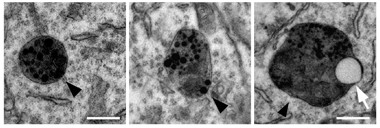
Compared to smooth lysosomes from wild-type mice (left), those from PLD3 knockouts (middle and right) are larger, dented, and may contain clear inclusions resembling lipid droplets (white arrow). [Fazzari et al., 2017. Nature.]
In 2013, Carlos Cruchaga of Washington University in St. Louis, and Alison Goate, now at Mount Sinai School of Medicine in New York, reported that two rare variants in the gene coding for PLD3, V232M and the synonymous A442A, double a person’s risk for AD (Dec 2013 news). They reported that overexpressing PLD3 reduced APP in N2A mouse neuroblastoma cells and lowered secretion of Ab42 and Ab40. Conversely, knocking down the protein bumped up Ab production. They also noticed brain levels of PLD3 being down in Alzheimer’s postmortem tissue. They hypothesized that PLD3 normally suppresses processing of APP.
Soon after, four papers called that association into question, reporting no, or only tenuous, links between V232M and late-onset disease (Apr 2015 news). A subsequent study did find an association of modest effect size in a study of Han Chinese (Zhang et al., 2016). De Strooper and colleagues decided to address the question mechanistically, exploring whether PLD3 affects APP processing.
To test the original claim, Fazzari and colleagues overexpressed wild-type PLD3 or the V232M mutant in HEK293 cells that also overexpressed wild-type human APP. As in the original study, cells overexpressing wild-type PLD3 secreted less Aβ into the medium than control cells, despite having normal levels of full-length APP. Curiously, though, Fazzari found that cells overexpressing the AD-associated PLD3 mutant also secreted less Aβ. According to Cruchaga’s analysis, this protein is a functional null that should have caused an uptick in Aβ. Could the APP and Aβ changes be an artifact of general PLD3 overexpression, which was 50 times greater than endogenous levels in all the cells, Fazzari wondered?
To check, the scientists created HEK293 cell lines that expressed 20, five, or only two times the endogenous amount of PLD3. At these levels, there was no difference in Aβ or full-length APP between cells expressing wild-type and mutant PLD3. The authors took this to mean that suppression of APP processing was an artifact of the 50-fold overexpression level.
To rule out an effect on APP processing, Fazzari examined PLD3 knockout mice. At three months, their cortices had the same levels of full-length APP, and soluble Aβ40 and Aβ42, as controls. When the researchers crossed PLD3 knockouts with APP knock-ins, they again saw no differences in APP processing or Aβ production in four-month-old animals compared to age-matched KI controls. Taken together, the data suggested that PLD3 does not affect APP processing in vitro or vivo. “We can say quite strongly say that the original hypothesis was incorrect,” Fazzari told Alzforum.
However, the researchers still found the genetic link to AD compelling. Was something else going on? Knowing that some AD-associated genes are implicated in endosomal–autophagic–lysosomal function, Fazzari examined these organelles in HEK293T cells. PLD3 co-localized with markers of late endosomes/lysosomes. When the researchers trained an electron microscope on these organelles from PLD3 knockout mice, they found them to be enlarged and shaped irregularly. They were stuffed with electron-dense inclusions—possibly packets of undigested waste—and many contained clear structures that could be lipid droplets, said Fazzari (see image above).
“This suggests that PLD3 loss impairs the function of the lysosome, and may render neurons more susceptible to accumulation of amyloid-β,” Fazzari told Alzforum. He added that this new hypothesis needs investigation. In addition, while he said these findings exclude a direct effect of PLD3 on APP cleavage and Aβ generation, the researchers still need to test aged mice to be sure there is no effect on plaques. It would also be interesting to cross the PLD3 knockouts with AD mice that model other aspects of the disease, such as those that express mutant tau, Fazzari said.
Is this another blow to PLD3 as a risk gene for Alzheimer’s? Scientists had varying opinions. “This paper from the De Strooper lab convincingly shows that there is no clear effect of PLD3 on APP biology and reignites the debate on whether PLD3 is an AD gene,” wrote John Hardy, University College London, to Alzforum. “One hopes that a clear outcome on PLD3 will come from the massive sequencing efforts both in the U.S. and in Europe.” The Alzheimer’s Disease Sequencing Project in the United States is conducting whole genome and exome sequencing studies on thousands of people, while in Europe a large consortium of researchers integrates exome sequencing data from AD cases and controls.
“They have done a well-controlled study,” said Goate. “It certainly argues the effect of these PLD3 mutations is not on amyloid accumulation.” However, Goate doesn’t think this study changes PLD3’s status as an AD gene candidate, especially given that other disease-associated genes have no direct impact on Aβ, either. “It argues that we as a field should be looking beyond Aβ accumulation as a means to understand the pathological effects of AD,” she said.
“This paper clearly excludes APP processing as a mechanism for PLD3,” said Alfredo Ramirez, University of Bonn, Germany. He found the lysosomal phenotype data interesting, but said it would be important to test whether the potential AD-associated V232M variant has any effect. It’s not yet clear, especially given the genetics data, whether this function of PLD3 is associated with susceptibility to the disease, he said.
Cruchaga agreed—based on this study and other recent evidence co-localizing PLD3 with lysosomal proteins—that the lipase could be acting through the lysosome (Satoh et al., 2014). He thinks this could have downstream consequences for APP processing and Aβ levels. Checking how dysfunctional lysosomes affect Aβ in aged APP mice will be important, said Celeste Karch, also at WashU.
Ralph Nixon, NYU School of Medicine, said this paper adds to accumulating evidence that the lysosome is important in AD and neurodegenerative disease and, if the genetic association bolds up, that it can be a cause, not just a downstream result of amyloid pathology. The changes in lysosomal structure reported in the current paper look similar to what researchers see in AD tissue from humans and animal models, he said, adding that it would be interesting to characterize the associated alterations as well.
"We are far from understanding PLD3’s definite role in the genetics and pathogenesis of disease,” wrote Cornelia van Duijn, Erasmus Medical Center, Rotterdam, The Netherlands, to Alzforum. “Fazzari and colleagues’ work underscores that functional studies of cellular models of genetic variants are error prone; independent replications are key.”—Gwyneth Dickey Zakaib
References
News Citations
- Phospholipase D3 Variants Double Sporadic AD Risk
- The PLD3 Gene: Alzheimer's Risk Factor or False Alarm?
Research Models Citations
Paper Citations
- Zhang DF, Fan Y, Wang D, Bi R, Zhang C, Fang Y, Yao YG. PLD3 in Alzheimer's Disease: a Modest Effect as Revealed by Updated Association and Expression Analyses. Mol Neurobiol. 2015 Jul 21; PubMed.
- Satoh J, Kino Y, Yamamoto Y, Kawana N, Ishida T, Saito Y, Arima K. PLD3 is accumulated on neuritic plaques in Alzheimer's disease brains. Alzheimers Res Ther. 2014;6(9):70. Epub 2014 Nov 2 PubMed.
Further Reading
Papers
- Zhang DF, Fan Y, Wang D, Bi R, Zhang C, Fang Y, Yao YG. PLD3 in Alzheimer's Disease: a Modest Effect as Revealed by Updated Association and Expression Analyses. Mol Neurobiol. 2015 Jul 21; PubMed.
- Wang C, Wang HF, Tan MS, Liu Y, Jiang T, Zhang DQ, Tan L, Yu JT, Alzheimer’s Disease Neuroimaging Initiative. Impact of Common Variations in PLD3 on Neuroimaging Phenotypes in Non-demented Elders. Mol Neurobiol. 2015 Aug 1; PubMed.
- Wang J, Yu JT, Tan L. PLD3 in Alzheimer's Disease. Mol Neurobiol. 2014 Jun 17; PubMed.
- Lambert JC, Grenier-Boley B, Bellenguez C, Pasquier F, Campion D, Dartigues JF, Berr C, Tzourio C, Amouyel P. PLD3 and sporadic Alzheimer's disease risk. Nature. 2015 Apr 2;520(7545):E1. PubMed.
- Satoh J, Kino Y, Yamamoto Y, Kawana N, Ishida T, Saito Y, Arima K. PLD3 is accumulated on neuritic plaques in Alzheimer's disease brains. Alzheimers Res Ther. 2014;6(9):70. Epub 2014 Nov 2 PubMed.
Primary Papers
- Fazzari P, Horre K, Arranz AM, Frigerio CS, Saito T, Saido TC, De Strooper B. PLD3 gene and processing of APP. Nature. 2017 Jan 25;541(7638):E1-E2. PubMed.
Annotate
To make an annotation you must Login or Register.

Comments
Institute of Neurology, UCL
This paper from the De Strooper lab convincingly shows that there is not a clear effect of PLD3 on APP biology and reignites the debate on whether PLD3 is an AD gene (Cruchaga et al., 2013).
Genetic analysis looking for rare variants is subject to difficult confounds. Usually, cases and controls are collected in subtly different ways and they may not be perfectly matched. In the U.K., for example, a higher proportion of controls were collected from the Cambridge area. These subtle population differences don’t cause a problem in GWAs with genes with modest effect sizes, but as studies look for GWAs hits of very small effect sizes or at rare variants like PLD3, these small population stratification issues can cause problems. For example, a founder variant in the Cambridge area could be mistaken for a protective allele in this sample. In the case of PLD3, replication attempts have given mixed results with no clear outcome (van der Lee et al., 2015; Lambert et al., 2015; Heilmann et al., 2015; Hooli et al., 2015). One hopes that a clear outcome on PLD3 will come from the massive sequencing efforts both in the U.S. and in Europe. PLD3 is not alone in remaining in pathogenic limbo. Since TREM2 was reported, no other new genes have yet been consistently replicated. In contrast, rare variants found in GWAs hits seem to give more consistent results.
Replication of genetic associations, outside of the first study, remains the most effective mechanism for deciding what is correct.
References:
Cruchaga C, Karch CM, Jin SC, Benitez BA, Cai Y, Guerreiro R, Harari O, Norton J, Budde J, Bertelsen S, Jeng AT, Cooper B, Skorupa T, Carrell D, Levitch D, Hsu S, Choi J, Ryten M, UK Brain Expression Consortium, Hardy J, Ryten M, Trabzuni D, Weale ME, Ramasamy A, Smith C, Sassi C, Bras J, Gibbs JR, Hernandez DG, Lupton MK, Powell J, Forabosco P, Ridge PG, Corcoran CD, Tschanz JT, Norton MC, Munger RG, Schmutz C, Leary M, Demirci FY, Bamne MN, Wang X, Lopez OL, Ganguli M, Medway C, Turton J, Lord J, Braae A, Barber I, Brown K, Alzheimer’s Research UK Consortium, Passmore P, Craig D, Johnston J, McGuinness B, Todd S, Heun R, Kölsch H, Kehoe PG, Hooper NM, Vardy ER, Mann DM, Pickering-Brown S, Brown K, Kalsheker N, Lowe J, Morgan K, David Smith A, Wilcock G, Warden D, Holmes C, Pastor P, Lorenzo-Betancor O, Brkanac Z, Scott E, Topol E, Morgan K, Rogaeva E, Singleton AB, Hardy J, Kamboh MI, St George-Hyslop P, Cairns N, Morris JC, Kauwe JS, Goate AM. Rare coding variants in the phospholipase D3 gene confer risk for Alzheimer's disease. Nature. 2014 Jan 23;505(7484):550-4. Epub 2013 Dec 11 PubMed.
van der Lee SJ, Holstege H, Wong TH, Jakobsdottir J, Bis JC, Chouraki V, van Rooij JG, Grove ML, Smith AV, Amin N, Choi SH, Beiser AS, Garcia ME, van IJcken WF, Pijnenburg YA, Louwersheimer E, Brouwer RW, van den Hout MC, Oole E, Eirkisdottir G, Levy D, Rotter JI, Emilsson V, O'Donnell CJ, Aspelund T, Uitterlinden AG, Launer LJ, Hofman A, Boerwinkle E, Psaty BM, DeStefano AL, Scheltens P, Seshadri S, van Swieten JC, Gudnason V, van der Flier WM, Ikram MA, van Duijn CM. PLD3 variants in population studies. Nature. 2015 Apr 2;520(7545):E2-3. PubMed.
Lambert JC, Grenier-Boley B, Bellenguez C, Pasquier F, Campion D, Dartigues JF, Berr C, Tzourio C, Amouyel P. PLD3 and sporadic Alzheimer's disease risk. Nature. 2015 Apr 2;520(7545):E1. PubMed.
Heilmann S, Drichel D, Clarimon J, Fernández V, Lacour A, Wagner H, Thelen M, Hernández I, Fortea J, Alegret M, Blesa R, Mauleón A, Roca MR, Kornhuber J, Peters O, Heun R, Frölich L, Hüll M, Heneka MT, Rüther E, Riedel-Heller S, Scherer M, Wiltfang J, Jessen F, Becker T, Tárraga L, Boada M, Maier W, Lleó A, Ruiz A, Nöthen MM, Ramirez A. PLD3 in non-familial Alzheimer's disease. Nature. 2015 Apr 2;520(7545):E3-5. PubMed.
Hooli BV, Lill CM, Mullin K, Qiao D, Lange C, Bertram L, Tanzi RE. PLD3 gene variants and Alzheimer's disease. Nature. 2015 Apr 2;520(7545):E7-8. PubMed.
Make a Comment
To make a comment you must login or register.