
T Cells from the Gut Occupy a Niche in Human and Mouse Brain
Quick Links
The brain and spinal cord were once thought to enjoy immune privilege, meaning they were believed to be largely insulated from immune activity elsewhere in the body. In recent years, however, researchers have discovered immune cells in various contexts throughout the central nervous system. Now, scientists led by David Hafler and Andrew Wang at Yale University in New Haven, Connecticut, have found a population of immune cells that travel to the brain from the gut or adipose tissue. In Nature May 28, they reported that these CD4-positive T helper cells take up residence in the subfornical organ, a structure that surveys the blood for nutrients and inflammatory markers. Without the interferon-γ secreted from these T cells, mice waited longer after a fast to begin eating, suggesting a role for this cell population in controlling feeding behavior.
- A cadre of T cells, originating in gut and fat tissue, resides in the brain.
- The cells sit in the subfornical organ, which surveils the blood.
- They affect food-seeking behavior by releasing interferon γ.
"This work takes us another step past the old dogma of CNS immune privilege and pushes us to think more broadly about immune-derived signals as physiological modulators of brain function,” wrote Jonathan Kipnis at Washington University in St. Louis (comment below).
Previously, the group discovered IFN-γ−secreting CD4+ T cells in cerebrospinal fluid samples from healthy people and from people with multiple sclerosis (Pappalardo et al., 2020). These findings hinted at a role for T helper cells in the central nervous system. To investigate whether such cells exist in the parenchyma, first author Tomomi Yoshida and colleagues immunostained mouse brain tissue for CD4+ T cells. The densest clusters occurred in the subfornical organ just under the hippocampus. Located near the third ventricle in the brain, the SFO is one of seven circumventricular organs, which lack complete blood-brain barriers. The SFO monitors the blood for inflammation and nutrition levels. The researchers found similar clusters of CD4+ T cells in the SFO of postmortem human brains.

T Cell Niche. The subfornical organ (SFO) sits under the fornix (fx) below the corpus callosum (cc). [Courtesy of Bentivoglio et al., Front Immunol., 2018.]
T cells have been found in the brain meninges as well (May 2021 news; Feb 2025 news), but flow cytometry and single-cell RNA sequencing confirmed that these SFO CD4+ T cells are distinct. They expressed higher levels of the cytokines IFN-γ and IL-17 as well as CXCR6, a chemokine receptor. Intriguingly, SFO T cell expression most closely resembled that of T cells from white adipose tissue.
Suspecting a connection between adipose tissue and SFO CD4+ T cells, Yoshida fed mice a high-fat diet to see if it changed both populations. As the mice packed on more weight, more CD4+ T cells occupied the SFO. To track if the brain T cells originate in white fat, the authors selectively labelled CD4+ T cells in adipose tissue. A day later, the highest levels of these labeled cells, outside of fat, were within the brain.
Because a high-fat diet can alter the microbiome, which in turn regulates T cell populations (Zheng et al., 2020), the authors wondered if the gut played a role in getting these T cells into the brain. Indeed, they found that CD4+ T cells only took hold in mice old enough to have a mature gut microbiome. Further, germ-free mice colonized with gut bacteria had more CD4+ T cells in the brain than counterparts without a gut microbiome. Once again, selective labeling indicated that CD4+ T cells made their way into the brain from the gut, suggesting the cells can come from both there and adipose tissue. Clonal overlap between T cells in the brain and those from fat and the gut further supported the notion that brain CD4+ T cells originate from those two sites.
How about in the human brain? Yoshida and colleagues analyzed gene-expression profiles of T cells from the blood, CSF, colon, and small intestine of a healthy donor. Cells from the CSF and small intestine shared common expression patterns, including high expression of IFN-γ and CXCR6 genes, suggesting a possible connection between gut and CNS T cells in people as well.
To test this connection in mice, they knocked out CXCR6. This reduced the number of CD4+ T cells in the brain parenchyma, but not in the meninges. In the SFO, high levels of the chemokine CXCL16, a ligand for CXCR6, supported that idea that this small part of the brain recruits T cells.
Once gut/adipose tissue T cells infiltrate the brain, do they have any physiological influence? Thinking the gut/adipose/brain connection might be important for control of appetite or foraging, the authors tested mice in a novelty-suppressed feeding task, which measures how long mice wait to eat after a fast if they are placed in a new environment. Compared to wild-type mice, T cell knockouts hesitated longer before eating. Transferring wild-type CD4+ T cells to these mice decreased waiting time, but not if the cells lacked IFN-γ, suggesting that food-seeking behavior is shaped by this interferon.
To investigate which cells are involved, the authors repeated their behavioral experiments on mice with IFN-γ receptors knocked out of microglia, neurons, or astrocytes. The latter waited more in the feeding task, just like mice lacking T cells, though for the IFN-γ receptor knockouts this was only true in males.
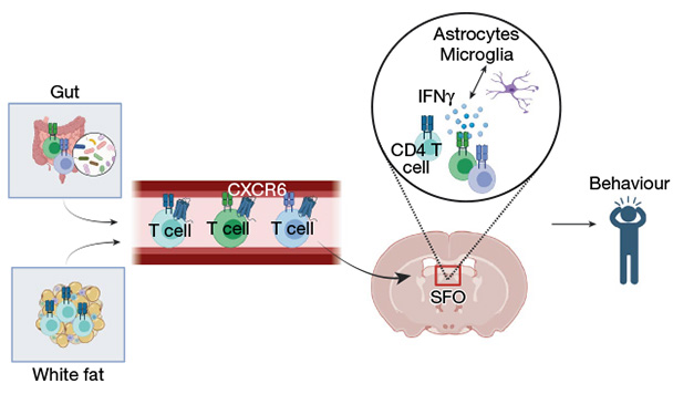
T Cell Troika. CD4+ T cells from the gut and white fat are guided by CXCR6 through the bloodstream into the SFO, where they help maintain homeostasis by releasing IFN-γ to drive food-seeking behavior. [Courtesy of Yoshida et al., Nature, 2025.]
These results indicate “a whole new role for the adaptive immune system” in the brain beyond disease, surveilling the bloodstream for nutritional information and influencing behavior accordingly, said Hafler. To him, the results point to a role for the immune system promoting physiological homeostasis in the brain. Still, he is investigating the relationship between this new gut-brain connection and neurological conditions such as Parkinson’s disease and multiple sclerosis.
Indeed, another study, published May 13 in the Journal of Clinical Investigation, offers more evidence for gut-driven immune influence on the brain—this time in Alzheimer's disease. Scientists led by Robert Vassar of the Northwestern University Feinberg School of Medicine in Chicago reported that the gut microbiome dictates IL-17 secretion from peripheral CD4+ T cells, and that this fosters amyloid plaques and astrocyte activation within the brain.
First author Sidhanth Chandra and collaborators built on their previous findings that mice treated with an antibiotic cocktail that slashed gut microbial diversity accumulated fewer plaques and had fewer activated astrocytes and microglia (Chandra et al., 2023). In the new study, they found that the antibiotics boosted levels of the short chain fatty acid propionate in the plasma of APP/PS1-21 mice. Administering just propionate to APP/PS1-21 mice reduced plaque load and soothed astrocytic activation.
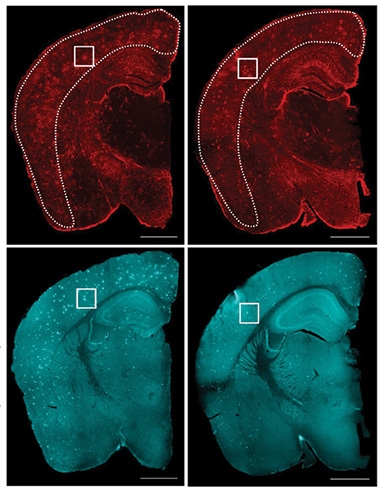
Appropriate Treatment? In APP/PS1-21 mice (left), astrocytosis (top) and amyloid plaques (bottom) were attenuated (right) by giving the animals proprionate for two months. [Courtesy of Chandra et al., Journal of Clinical Investigation, 2025.]
How does a compound secreted by gut bacteria exert influence on the brain? Flow cytometry experiments indicated that propionate reduced the Th17 cell population in tissue samples from various mouse organs, as well as the IL-17 secreted from these cells. An antibody against IL-17 decreased plaque load and astrocyte activation about as well as IL-17 antibody and propionate treatment combined, indicating that the cytokine is a key link between gut bacteria and astrocytosis. The researchers are studying the mechanisms involved.
These results suggest that “astrocytes are not passive bystanders but active integrators of microbial and immune signals, capable of shaping CNS pathology,” wrote Rudolph Tanzi and Nanda Kumar Navalpur Shanmugam of Harvard University (comment below).
“It's becoming clearer that there's interplay between the innate immune system, microglia, and astrocytes, but also between the gut microbiome and the adaptive immune system,” said Vassar. “That's a new understanding that we didn't have before, and we're hoping to exploit it therapeutically to help people with Alzheimer's.”—Lauren Schneider
Lauren Schneider is a freelance writer in New York City.
References
News Citations
- As Mice Age, T Cells Traipse Around Their Meninges. Mayhem Ensues
- From the Meninges, Regulatory T Cells Keep Brain Inflammation in Check
Paper Citations
- Pappalardo JL, Zhang L, Pecsok MK, Perlman K, Zografou C, Raddassi K, Abulaban A, Krishnaswamy S, Antel J, van Dijk D, Hafler DA. Transcriptomic and clonal characterization of T cells in the human central nervous system. Sci Immunol. 2020 Sep 18;5(51) PubMed.
- Zheng D, Liwinski T, Elinav E. Interaction between microbiota and immunity in health and disease. Cell Res. 2020 Jun;30(6):492-506. Epub 2020 May 20 PubMed.
- Chandra S, Di Meco A, Dodiya HB, Popovic J, Cuddy LK, Weigle IQ, Zhang X, Sadleir K, Sisodia SS, Vassar R. The gut microbiome regulates astrocyte reaction to Aβ amyloidosis through microglial dependent and independent mechanisms. Mol Neurodegener. 2023 Jul 6;18(1):45. PubMed.
Further Reading
Papers
- Pappalardo JL, Zhang L, Pecsok MK, Perlman K, Zografou C, Raddassi K, Abulaban A, Krishnaswamy S, Antel J, van Dijk D, Hafler DA. Transcriptomic and clonal characterization of T cells in the human central nervous system. Sci Immunol. 2020 Sep 18;5(51) PubMed.
- Chandra S, Di Meco A, Dodiya HB, Popovic J, Cuddy LK, Weigle IQ, Zhang X, Sadleir K, Sisodia SS, Vassar R. The gut microbiome regulates astrocyte reaction to Aβ amyloidosis through microglial dependent and independent mechanisms. Mol Neurodegener. 2023 Jul 6;18(1):45. PubMed.
- Zheng D, Liwinski T, Elinav E. Interaction between microbiota and immunity in health and disease. Cell Res. 2020 Jun;30(6):492-506. Epub 2020 May 20 PubMed.
- Bentivoglio M, Kristensson K, Rottenberg ME. Circumventricular Organs and Parasite Neurotropism: Neglected Gates to the Brain?. Front Immunol. 2018;9:2877. Epub 2018 Dec 11 PubMed.
Primary Papers
- Yoshida TM, Nguyen M, Zhang L, Lu BY, Zhu B, Murray KN, Mineur YS, Zhang C, Xu D, Lin E, Luchsinger J, Bhatta S, Waizman DA, Coden ME, Ma Y, Israni-Winger K, Russo A, Wang H, Song W, Al Souz J, Zhao H, Craft JE, Picciotto MR, Grutzendler J, Distasio M, Palm NW, Hafler DA, Wang A. The subfornical organ is a nucleus for gut-derived T cells that regulate behaviour. Nature. 2025 May 28; Epub 2025 May 28 PubMed.
- Chandra S, Popovic J, Singhal NK, Watkins EA, Dodiya HB, Weigle IQ, Salvo MA, Ramakrishnan A, Chen Z, Watson JT, Shetti A, Piehl N, Zhang X, Cuddy LK, Sadleir KR, Schwulst SJ, Prakriya M, Gate D, Sisodia SS, Vassar R. The gut microbiome controls reactive astrocytosis during Aβ amyloidosis via propionate-mediated regulation of IL-17. J Clin Invest. 2025 May 13; Epub 2025 May 13 PubMed.
Annotate
To make an annotation you must Login or Register.
Comments
Washington University in St. Louis, School of Medicine
This is a beautiful and important study. It adds a new layer to our understanding of how the immune system communicates with the brain under homeostatic conditions. The idea that gut-primed T cells migrate to the subfornical organ and shape behavior via IFN-γ resonates strongly with our earlier findings (Filiano et al., 2016), where we showed a role for IFN-γ in regulating social behavior.
What’s exciting here is the clear identification of brain-resident T cells—not just in the meninges—and the evidence that they are transcriptionally distinct, CXCR6-positive, and behaviorally relevant. This work takes us another step past the old dogma of CNS immune privilege and pushes us to think more broadly about immune-derived signals as physiological modulators of brain function.
References:
Filiano AJ, Xu Y, Tustison NJ, Marsh RL, Baker W, Smirnov I, Overall CC, Gadani SP, Turner SD, Weng Z, Peerzade SN, Chen H, Lee KS, Scott MM, Beenhakker MP, Litvak V, Kipnis J. Unexpected role of interferon-γ in regulating neuronal connectivity and social behaviour. Nature. 2016 Jul 21;535(7612):425-9. Epub 2016 Jul 13 PubMed.
Massachusetts General Hospital
Massachusetts General Hospital/Harvard Medical School
Chandra et al. have demonstrated novel pathways through which the gut microbiome modulates brain pathology in Alzheimer’s disease. They also propose molecular mechanisms that could be targeted for therapeutic intervention. The highlight of this elegant study is its careful dissection of astrocytic reactivity in the context of Aβ pathology. While prior studies have mainly focused on microglial responses or amyloid deposition, Chandra et al. demonstrate a role for astrocytes in responding to gut-derived cues, most notably the short-chain fatty acid (SCFA), propionate.
Plasma metabolomics revealed a selective increase in propionate in antibiotic-treated mice, which negatively correlated with markers of astrocyte reactivity and positively with homeostatic astrocyte traits. Importantly, this increase was linked to elevated abundance of Akkermansia muciniphila, a gut commensal known to produce propionate. This association draws attention to the specific microbial contributors to neuroactive metabolite pools and highlights the potential of targeting select bacterial taxa to therapeutically modulate factors that cause brain inflammation.
The authors also show that direct administration of sodium propionate recapitulates the effects of broad-spectrum antibiotic treatment, suppressing astrocytic reactivity and reducing Aβ plaque burden—while sparing microglial function. These results argue that astrocytes are not passive bystanders but active integrators of microbial and immune signals, capable of shaping CNS pathology. Importantly, these neuroinflammatory changes were accompanied by increased levels of synaptic proteins, including synaptophysin and PSD-95, with propionate treatment. This suggests that reducing astrocytic reactivity may preserve, or even enhance, synaptic integrity—a vital consideration given the strong association between synapse loss and cognitive decline in AD. This finding adds a new layer to our understanding of how the gut microbiome can impact neurodegenerative processes beyond the realm of immune activation and amyloid deposition.
By linking propionate exposure to the suppression of peripheral Th17 cell differentiation and IL-17A production, the authors also identify a novel immune-metabolic axis that bridges gut microbial metabolites with CNS inflammation. Using IL-17A monoclonal antibody treatment, they show that the effects of propionate on reducing astrocyte reactivity and Aβ pathology are IL-17-dependent. These experiments underscore how modulation of a single metabolite—propionate—can critically influence CNS inflammatory pathways via T-cell mediated immune responses.
From a translational perspective, the identification of a naturally occurring microbial metabolite that ameliorates AD pathology opens new therapeutic possibilities. Propionate—or microbial consortia and dietary strategies designed to enhance SCFA production—could represent a new class of adjunct therapies aimed at modifying disease progression by targeting gut-CNS communication.
This study challenges the microglia-centric view of AD by highlighting astrocytes as independent, microbiota-responsive immune players, urging a more integrated perspective that bridges immunology and neurobiology.
In conclusion, Chandra et al. present a conceptually bold and methodologically rigorous study that advances our understanding of how peripheral systems influence CNS pathology in Alzheimer’s disease. Their identification of the propionate–IL-17–astrocyte axis represents an important advance in AD research. Future studies inspired by this work could explore how microbial ecology, dietary fiber intake, and targeted microbiome manipulation intersect with host immunity to affect brain aging and resilience to Alzheimer’s disease and other neurodegenerative disorders.
Make a Comment
To make a comment you must login or register.