
Killers in the Crosshairs: Zeroing in on Invading CD8+ T Cells
Quick Links
As the trained killers of the immune system, cytotoxic T cells silently save our lives every day—vanquishing viruses before they can run amok, and putting nascent cancer cells out of business before they set up shop. But when these powerful responses are misguided, they can do harm. Case in point: Evidence has been mounting in recent years that brain-infiltrating T cells play a hand in neurodegeneration, including in Alzheimer’s and Parkinson’s diseases. They are of the cytotoxic type. What beckons them to the brain in the first place? Findings presented at the AD/PD meeting, held earlier this month in Vienna, illuminated some possible mechanisms.
- In tauopathy mice, conventional dendritic cells (cDC) prime cytotoxic T cells to maraud the brain.
- These cDCs present brain-derived antigens to T cells sitting in the cervical lymph nodes.
- In ApoE4 carriers, CD8+ T cells more easily cross from the blood into the brain.
In tauopathy mice, antigens from the brain can drain via dural lymphatics into deep cervical lymph nodes, where a contingent of resident dendritic cells gobble them up and present them to T cells. Once primed to the scent of their prey, these cells then enter the brain to seek and destroy. Intriguingly, in ApoE4 carriers, killer T cells more readily cross into the brain. Researchers are developing therapeutics to thwart the advances of these misbegotten cells, and they showed results from a handful of small trials in Vienna (see Part 13 of this series).
This focus on targeting T cells is part of a broader movement directed at the neuroinflammatory aspects of Alzheimer’s and related diseases. Other immune-based approaches include supporting microglia by turning on TREM2 (see Part 5 of this series) or inhibiting checkpoint proteins that hold microglia back (see upcoming Alzforum story).
The healthy brain hosts very few T cells. However, they can be summoned to the brain when threats arise. This seems to be the case in people with AD or PD, whose CSF and brain tissue swarm with CD8+ T effector memory cells (Jan 2020 news). This type of T cell has been previously primed, specifically via MHC I molecules that present antigens that have been processed inside the cell. Once the T cells have recognized their designated antigen and clonally expanded in response, legions of them will kill any cell presenting said antigen. Such murderous cells were spotted cavorting with microglia around plaques in brain samples from people with AD (Dec 2022 news). A glut of primed-and-ready CD8+ T cells has also been found circulating in the blood of people with AD (Feb 2024 news).
Meanwhile, David Holtzman of Washington University in St. Louis identified T cells as troublemakers in mouse models of tauopathy. In ApoE4/tau transgenics, distressed microglia somehow beckoned CD8+ T cells into the brain, and removing these infiltrators halted neurodegeneration (Mar 2023 news).
What draws these T cells into the brain? After all, killer T cells aren’t turned on willy-nilly—they only become activated when their T cell receptor engages its cognate antigen. What’s more, to turn on a naïve T cell for the first time, the antigen must be presented by a professional antigen-presenting cell (APC) equipped with co-stimulatory bells and whistles. Where in the body, and by what APCs, are these antigens being flaunted? In Vienna, Holtzman presented new data that chip away at this pivotal question.
Dendritic cells (DCs) are highly specialized APCs. Some are stationed in various tissues throughout the body. Others, called conventional DC 1, reside in lymph nodes, where they continually phagocytose antigens draining there from nearby tissues, process them internally, and present them to T cells. The brain has nary any of these cDC1s. Still, Holtzman speculated that cDC1s might be responsible for priming the CD8+ T cells that would later invade the brain. To find out, postdoc Hao Hu generated mice missing the Irf8Δ+32 enhancer, which is essential for cDC1 development.
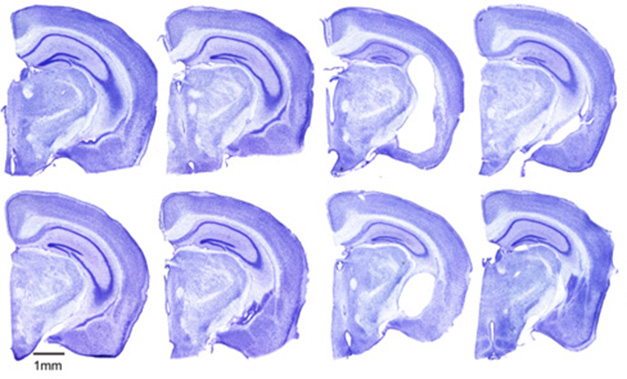
Powerful Protection. Compared with human ApoE4 wild-type (left column) or human ApoE4 cDC1-deficient (center left) mice, mutant tau transgenic ApoE4 mice (center right) have profound neurodegeneration, as seen by tissue loss and enlarged lateral ventricles. Deleting cDC1 cells in these mice (right column) spares neurons. Males (top) have more shrinkage than females (bottom). [Courtesy of David Holtzman, Washington University, St. Louis, 2025.]
Nixing cDC1s from tau/ApoE4 transgenics (TE4) in this way did not influence the extent of their tau pathology. Instead, it put a wrench in its downstream neurodegenerative cascade—preventing activation of microglia and astrocytes, stemming a rise in plasma NfL, and protecting the hippocampus from shrinkage (image above). Holtzman said that this protection was just like what his group had seen previously when they removed T cells. Indeed, removal of cDCs halted the infiltration of CD8+ T cells into the brains of TE4 mice. It left unchanged the number of CD4+ T cells entering the brain. Curiously, though, it enhanced infiltration of natural killer (NK) and NKT cells (more on NK cells later).
Holtzman said that these experiments have not yet been done in tauopathy mice expressing ApoE3 instead of E4. He suspects that the same T-cell cascade is likely instigated in response to tauopathy regardless of ApoE genotype. ApoE4 just makes everything worse, as reported previously (Sep 2017 news).

Presentation Station. Within deep cervical lymph nodes, pictured here in a mouse, cDC1 cells internalize antigens draining from the meningeal lymphatic vessels and present them to CD8+ T cells. The primed T cells then infiltrate the brain. [Courtesy of Hao Hu, Washington University, St. Louis, 2025.]
If not within the brain itself, where were these cDCs priming CD8+ T cells? Because brain-derived antigens flow via the dural lymphatics into the deep cervical lymph nodes (dCLN) positioned just outside of the brain, Holtzman reasoned that this might be where CD8+ T cells were receiving their marching orders. To find out if this might be the case, the scientists transfected MHC I knockout mouse neurons with the antigenic protein ovalbumin; this equipped the cells with antigen without the machinery to present it to T cells. They then injected these cells into the brains of wild-type mice. The scientists also intravenously injected the mice with OT-1 T cells—an ovalbumin-specific CD8+ T cell clone. A week later, the scientists found that, indeed, these Ova-specific T cells had expanded within the cervical lymph nodes (and in the spleen). Importantly, deletion of cDC1 cells completely nixed this expansion. Together, this suggested that cDC1 cells stationed within the dCLNs prime CD8+ T cells with brain-derived antigens.
Holtzman emphasized to Alzforum that these experiments only demonstrate that cDCs stationed in the dCLNs are capable of presenting brain-derived antigens to CD8+ T cells. Whether this route is how CD8+ T cells get primed in neurodegenerative disease remains unknown, as does the identity of those antigens. He speculates that, in the context of AD, where neurons start churning out phosphorylated forms of tau that the immune system never encountered during development, perhaps snippets of this modified tau appear foreign, riling T cells in the dCLNs that happen to express a TCR that recognizes them. It’s also possible that T cells are activated by other antigens released from damaged or dying neurons, he suggested.
Regarding this Alzheimer’s context, Sem Halters of Amsterdam University Medical Center made the case that ApoE4 eases the passage of antigen-primed CD8+ T cells into the brain. Halters, a graduate student in the lab of Helga de Vries, looked for differences in the composition of peripheral immune cells in human-ApoE targeted replacement mice. In short, he found that mice expressing ApoE4 had fewer naïve and effector CD8+ T cells, but more memory CD8+ T cells in their spleens, than did ApoE3 mice. “This is of critical importance, because it is these memory CD8+ T cells that are thought to infiltrate the brain in AD patients,” Halters said.
Halters reported that relative to CD8+ T cells from ApoE3 mice, those isolated from ApoE4 mice expressed more LFA-1. This receptor binds ICAM1 on the brain’s endothelial cells, which facilitates transendothelial migration of immune cells into the brain. In cell culture assays, ApoE4 CD8+ T cells wriggled across a layer of brain endothelial cells more efficiently than did their E3 counterparts. In the brains of 6-month-old mice, Halters noticed that regardless of their ApoE genotype, he found more CD8+ T cells in white matter than in gray matter, though in white-matter tracts such as the corpus collosum, CD8+ T cells were much more abundant in E4 than E3 mice.
This held true in human postmortem brain samples, where Halters found twice as many CD8s within white matter of E4/E4 carriers with AD than in their E3/E3 counterparts. White matter of non-demented controls housed fewer CD8s still.
Furthermore, microglia residing within white matter expressed a more activated profile in E4/E4 carriers. Halters does not know whether the infiltrating CD8s rile up the microglia, or if riled-up microglia summon CD8s to the white matter. It could also go both ways, he said. While Halters is still piecing together the mechanisms underlying these effects, he thinks something about the activation of the brain endothelium in ApoE4 carriers seems to play a hand in easing the entry of CD8+ T cells.
Some amount of consensus seems to be building that CD8+ T cells summoned to the brain are up to no good in the context of neurodegenerative disease. However, as with all things immunological, the story is not simple. For example, one study led by Hongbo Chi at St. Jude Children’s Hospital in Memphis, Tennessee, cast a population of brain-resident CD8+ T cells in a beneficial light (Su et al., 2023). Dubbed TRM, these cells expressed the chemokine receptor CXCR6, and hobnobbed with plaque-associated microglia in the 5xFAD mouse model of amyloidosis, where they supported microglial removal of plaques. These findings appear to contradict the picture that Holtzman, Gate, and other scientists have painted about the cells.
Holtzman noted important differences between these studies. For one, his and other groups have found that infiltrating CD8+ T cells exacerbate tau-mediated neurodegeneration, whereas Chi’s study used an amyloidosis model, which does not undergo substantial neuronal loss. For another, the numbers of brain-resident TRM cells found in Chi’s study paled in comparison to the hordes of infiltrators found in the tauopathy models.
“The T cells that are in the brain in the tauopathy model are very likely coming from outside the brain and are not likely ‘TRM’ cells,” Holtzman told Alzforum. “While Su et al. suggests that CXCR6+CD8+ T cells and IFN-γ may exhibit protective roles in mouse models that only have amyloid pathology, other studies indicate that T cells are more closely associated with tau pathology and promote neurodegeneration,” he added.
It’s possible that, akin to what scientists have observed about the microglial receptor TREM2, the role of CD8+ T cells may shift depending on the neuropathology at hand—which itself evolves as this long disease progresses over the course of years.
Even so, scientists are moving forward with small trials that aim to disarm purportedly cytotoxic T cells in AD and other neurodegenerative diseases. See next story for more about these first attempts.—Jessica Shugart
References
News Citations
- Therapies Aim to Tame T Cells in the Brain
- Trialists Grapple with How to Outsmart TREM2
- Attack of the Clones? Memory CD8+ T Cells Stalk the AD, PD Brain
- In AD, CSF Immune Cells Hint at Mounting Mayhem in the Brain
- Epigenetic Shenanigans—In AD, Chromatin Opens Up in Blood Immune Cells
- Neurodegeneration—It’s Not the Tangles, It’s the T Cells
- ApoE4 Makes All Things Tau Worse, From Beginning to End
Paper Citations
- Su W, Saravia J, Risch I, Rankin S, Guy C, Chapman NM, Shi H, Sun Y, Kc A, Li W, Huang H, Lim SA, Hu H, Wang Y, Liu D, Jiao Y, Chen PC, Soliman H, Yan KK, Zhang J, Vogel P, Liu X, Serrano GE, Beach TG, Yu J, Peng J, Chi H. CXCR6 orchestrates brain CD8+ T cell residency and limits mouse Alzheimer's disease pathology. Nat Immunol. 2023 Oct;24(10):1735-1747. Epub 2023 Sep 7 PubMed.
Further Reading
Annotate
To make an annotation you must Login or Register.
Comments
No Available Comments
Make a Comment
To make a comment you must login or register.