TfR, No Longer Lone Star? New Shuttles Use Other Keys to Unlock Brain
Quick Links
The highly selective border lining the brain’s vasculature is the Achilles’ heel of any large therapeutic molecule aiming for targets in the brain, anti-Aβ antibodies included. Not only do fewer than 1 percent of these molecules manage to cross from the blood into the brain, but those that do tend to get snagged by clumps of vascular amyloid just on the other side, where they can instigate inflammation and dangerous brain swelling (e.g., Aug 2023 conference news).
- Denali’s tweaked, TfR-based shuttle gets anti-Aβ antibodies to brain plaques without harming reticulocytes.
- Dual shuttles aimed at both TfR and CD98hc maximize brain uptake and exposure relative to either alone.
- Grabody B is an IGFR1-based shuttle. It reportedly co-opts an actin-based internalization pathway to quickly transport antibody cargo.
Active transport strategies, such as transferrin receptor binding regions, circumvent these problems, ferrying their attached cargo across the brain’s capillaries and into the parenchyma. Trontinemab—a bispecific antibody that recognizes both Aβ and TfR—is farthest along in Alzheimer’s disease trials, where it has been shown to wipe out plaques throughout the brain while triggering minimal brain swelling, aka ARIA (see Part 9 of this series). Along with these clinical findings, new preclinical data published in Science and presented at this year’s AAIC held July 27-31 in Toronto, suggest that trontinemab and other TfR-based shuttles are no longer the only game in town.
Alternatives include Denali’s ATVcisLALA:Aβ, which uses a modified version of bispecific Aβ/TfR antibody with dampened effector function, allowing the antibody to engage its target in the brain without harming TfR-expressing reticulocytes in the blood. Besides co-opting the iron-transporting TfR to do their bidding, Denali and other groups are investigating the shuttling capabilities of CD98hc, a brain endothelial receptor that helps transport amino acids into the brain. According to findings presented at the conference, CD98hc-targeted shuttles ferry their cargo into the brain more slowly than TfR-targeted counterparts. Dual shuttles targeting both TfR and CD98hc appear to tap the best of both worlds, getting cargo into the brain faster and more extensively than either shuttle alone. Finally, a shuttle called Grabody B, which uses the lesser-known candidate insulin growth factor receptor (IGFR1), reportedly sneaks its cargo into the brain via a quick, actin-dependent endocytosis pathway to deliver therapeutic antibodies into the brain.
“This is an extremely exciting time for the explosion of BBB-enabled technology development and the first clinical proof-of-concept that is now emerging,” Joy Zuchero of Denali told Alzforum, adding “All the preclinical research in this space holds significant promise to improve the next generation of CNS drugs for both AD as well as other neurological diseases.” Zuchero co-chaired a session focusing on active transport mechanisms at AAIC.
For her part, Zuchero presented findings on Denali’s new and improved antibody transport vehicle, ATVcisLALA, most of which Alzforum covered previously as reported in a preprint (Oct 2024 news). The work appeared in Science on August 7 (Pizzo et al., 2025).
In brief, Zuchero and colleagues improved their original ATV. This ATV is a monoclonal antibody with a TfR-binding region inserted into one of its Fc regions (image below). In mouse models, an Aβ-targeted version of this ATV crosses into the brain and latches onto Aβ plaques in the parenchyma. However, as with other TfR-binding shuttles, it also binds to TfR-expressing reticulocytes in the blood and can derail their development into red blood cells.
To counteract this problem, the scientists introduced LALA mutations, which quash antibody effector function, into one or both Fc regions of the ATV. They found that when introduced into the same Fc arm that carries the TfR binding domain—the cisLALA configuration—the mutations extinguished effector function against reticulocytes, while leaving the anti-Aβ response intact. In contrast, reticulocyte targeting continued unabated when the LALA mutations were introduced into the opposing Fc arm (trans-LALA). Rendering both Fc arms effectorless went too far, dampening the antibody’s attack against Aβ.
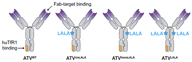
The LALA Compromise. The wild-type ATV features a TfR binding region on one Fc arm (left). Introducing the LALA mutation into the same arm (cisLALA) but not the opposing arm (transLALA) prevents damage to TfR-expressing reticulocytes. Adding LALA to both arms also extinguishes effector function against the target of the Fab arms. [Courtesy of Pizzo et al., Science, 2025.]
The published paper includes some additional experiments beyond those in the preprint. Notably, first author Michelle Pizzo and colleagues tested the limits of ATVcisLALA’s hematological safety by injecting mice with 13 weekly doses of the antibody, as opposed to a single injection. Even with this chronic regimen, they found that ATVcisLALA:Aβ had no impact on reticulocytes, red blood cell count, or hemoglobin levels.
In Toronto, Zuchero reported more of the main findings from the study. The highlights? When injected intravenously into 5xFAD mice expressing a humanized TfR, ATVcisLALA:Aβ crossed into the brain five- to eightfold more efficiently than an unmodified Aβ antibody. There, it was broadly distributed throughout the brain parenchyma, where it clung to plaques and summoned microglia to the scene. Adding both the TfR binding region and the cisLALA mutations to 3D6—the murine version of notorious ARIA instigator bapineuzumab—lowered the incidence of ARIA-like lesions and the intensity of vascular inflammation in mice.
Zuchero attributes this reduction in vascular pathology to ATVcisLALA:Aβ’s passage across capillaries, rather than at larger vessels where vascular amyloid tends to accumulate. In support of this, an unpublished light sheet microscopy image Zuchero shared with Alzforum shows an unmodified anti-Aβ antibody glommed onto vascular amyloid deposits lining large brain surface vessels in a 5xFAD mouse, whereas ATVcisLALA:Aβ is scarce, having crossed into the parenchyma via tiny vessels deeper in the brain instead.

Fuel for ARIA? Light sheet microscopy comparison between distribution of unmodified anti-Aβ (left) and ATVcisLALA:Aβ (right) along large vessels near the brain surface. While anti-Aβ associated with vascular amyloid deposits (yellow, co-localization), ATVcisLALA:Aβ sidestepped this interaction. [Courtesy of Meredith Calvert, Denali Therapeutics.]
A Perfect Pair? TfR and CD98hc
Despite this promising data, TfR-based shuttles aren’t the end all, be all, Zuchero acknowledged. Case in point: In another recent paper published July 22 in Cell Reports, her team introduced a dual ATV, equipped with both TfR and CD98hc binding domains. Like TfR, CD98hc is highly expressed on brain endothelial cells. The scientists previously reported that it, too, can ferry hefty therapeutics into the brain (Zuchero et al., 2016; Chew et al., 2023). However, the transport kinetics differ markedly between the two. TfR is a hare, rapidly whisking its cargo through brain endothelial cells and into the parenchyma, where it is, however, promptly internalized and catabolized by neurons on the other side. In contrast, CD98hc is more of a tortoise, taking days to transport its cargo into the brain, where it then sticks around for much longer than TfR-transported cargo does.
As reported in the new study, co-led by Zuchero, Kylie Chew, and Robert Wells of Denali, putting the two transporters together yields the best of both. That is, a dual ATV equipped with both TfR and CD98hc binding domains enters the brain faster than an ATV that only binds CD98hc, and is retained there for longer than one that only binds TfR (image below).
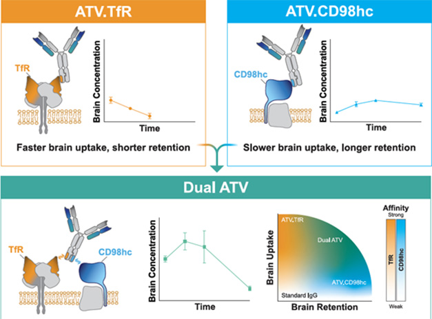
Green Light. TfR-based ATVs quickly cross into the brain but don’t last (top left). CD98hc-based ATVs get in more slowly, but stay longer (top right). Combining both yields the highest overall exposure (bottom). [Courtesy of Wells et al., Cell Reports, 2025.]
Zuchero did not show findings from this dual ATV study at AAIC. However, Liqun Wang of Harvard University’s Wyss Institute presented results using a similar dual shuttle approach. Wang’s brain shuttle platform makes use of nanobodies. Also known as VHH fragments, nanobodies are produced by immunized llamas. Wang generated nanobodies against both TfR and CD98hc, which could then be attached, either alone or in combination, to a monoclonal antibody cargo, or to other therapeutic molecules such as enzymes or antisense oligonucleotides. For these experiments, Wang attached anti-BACE1 antibodies as cargo.
In Toronto, Wang reported similar differences in the kinetics of brain uptake between the two transport vehicles as Zuchero published: namely, that TfR-based shuttles triggered rapid transport of their antibody cargo, whereas the CD98hc-targeted variety facilitated a slower passage into the brain, followed by prolonged exposure.
A closer look at where the shuttles went following injection revealed different distribution patterns, as well. While the TfR-specific nanobody shipped its antibody cargo into the brain on day 1 and promptly co-localized with neurons, the CD98hc shuttle lingered around brain capillaries for a week or so before crossing into the brain, where it spread out diffusely instead of associating with a specific cell type.
Finally, Wang reported that adding both types of nanobody shuttle to an anti-BACE1 antibody had a synergistic effect, leading to higher overall brain exposure than either one alone.
IGF1R Shuttle—Speedier Than TfR?
Insulin-like growth factor 1 receptor (IGF1R) has also emerged as a brain shuttle contender. In Toronto, Sungwon An of ABL Biosciences in South Korea presented fresh data on Grabody B, a therapeutic antibody shuttle with a single-chain variable fragment (scFv) specific for IGF1R attached to one of its Fc regions. Both Fc arms of this shuttle are engineered to enhance its plasma stability (image below).

Grab That IGF1R. The Grabody B brain shuttle features an anti-IGF1R antibody fragment fused to a therapeutic antibody of choice, along with engineered Fc regions to boost plasma stability. [Courtesy of Sungwon An, ABL Biosciences.]
IGF1R is highly expressed by brain capillary endothelial cells, as well as neurons and other cell types within the brain. Previous studies equipping an α-synuclein antibody with the Grabody B shuttle had demonstrated that the shuttle boosted the antibody’s passage into the brain by nearly an order of magnitude, and left normal signaling through IGF1R untouched. In a mouse model of Parkinson’s disease, the “shuttled” version of the antibody outperformed a conventional α-synuclein antibody in reducing α-synuclein aggregates, sparing neurons, and rescuing behavioral deficits (Shin et al., 2022). After promising preclinical data in nonhuman primates (An et al., 2024), a clinical version of this molecule—ABL301—was evaluated in a Phase 1 clinical trial in collaboration with Sanofi.
In Toronto, An reported that Grabody B has a distinct modus operandi to usher its cargo across the brain endothelium. She treated cultured human brain endothelial cells with BACE1 antibodies fused with TfR, CD98hc, or Grabody B (IGFR-1)-targeted shuttles. Grabody B left both the tortoise and the hare in the dust. More like a cheetah, Grabody B was internalized and appeared within intracellular puncta within 30 seconds, a process that took 30-120 minutes for the other two. Both Grabody B- and TfR-based shuttles gathered in vesicular puncta inside of cells, whereas CD98hc shuttle had a more thread-like distribution.
Further experiments revealed that while TfR-mediated internalization uses a classic clathrin-mediated endocytosis, the IGF1R-based Grabody B shuttle takes a different route. It goes across using both clathrin and caveolin. (Caveolin transcytosis of fibrinogen has been implicated in vascular amyloid deposition (Jul 2025 news). In An’s study, high-resolution microscopy showed that while the TfR shuttle co-localized strongly with early endosomes, Grabody B instead clung to actin filaments (image below).

Actin Tightrope Act. An antibody fused to Grabody B (green) associated with actin filaments (yellow arrows, co-localization). Under higher resolution, the brain shuttle appears to “hang” onto actin. [Courtesy of Sungwon An, ABL Biosciences.]
These characteristics suggested that Grabody B may be internalized via the lesser-known Fast Endophilin Mediated Endocytosis (FEME) pathway. Mediated by actin reorganization, this pathway is lightning-fast compared to clathrin-mediated endocytosis. Further work supported this hypothesis, as the scientists found that Grabody B co-localized not only with filamentous actin, but also with endophilin A2, another component of the FEME pathway. TfR-based shuttles associated with neither of these. Finally, the scientists spotted endophilin A2 lining the brain microvasculature in 5xFAD mice, and found that a BACE1 antibody equipped with Grabody B, but not one affixed with a TfR-based shuttle, co-localized with this actin-associated protein on the surface of vessels.
Benjamin Wolozin of Boston University asked the presenters how selective these new shuttle targets were for the brain endothelium, as opposed to other organs or cells in the body. Wang and other presenters acknowledged that all of the shuttles identified so far have some degree of expression in peripheral tissues. However, Wang noted that whether off-target binding of the shuttle causes peripheral side effects will depend on whether the target of its therapeutic cargo is also expressed in that same location. As more shuttle options are validated, scientists may be able to choose the right shuttle/therapeutic combination to minimize off-target effects.
The Boston-based biotech company Manifold Bio is attempting to generate a panoply of brain shuttles for scientists to choose from. As reported by Manifold’s Kimberly Scearce-Levie at AAIC, the company’s approach combines iterative rounds of AI-guided protein design followed by in vivo multiplexing of hundreds of potential shuttle proteins. So far, this has yielded several hundred proteins that could make brain shuttles for different types of targets, Scearce-Levie said. She reported that one of their candidates—dubbed PX1—was more specifically and highly expressed on brain endothelial cells than either TfR or CD98hc. When attached to an anti-Aβ antibody, it improved the antibody’s passage into the brain and its decoration of Aβ plaques in a 5xFAD mouse model.
Scearce-Levie would not reveal the identity of the candidate shuttle protein. Later, in response to a question, she acknowledged that PX1 is expressed in peripheral tissues, including liver and lung, but that it did not appear to cause pathology in either of those organs. “I think it does speak to this point that pure specificity is very difficult to achieve, but having a pattern of selectivity that can widen your therapeutic window is really the goal that we’re all trying to achieve,” Scearce-Levie said.
One attendee asked whether it might be more productive to focus on maximizing delivery and exposure of shuttle targets in the brain, rather than getting hung up on brain specificity. Zuchero responded that it is important to keep looking for shuttle targets that are as specific as possible and also boost brain exposure of their cargo—as both her and Wang’s data with dual TfR/CD98hc shuttles suggest is possible. However, she thinks brain shuttle science is shifting. “For a long time, the field was focused on increasing exposure as the benchmark for what makes a good transporter,” Zuchero said. “Now we’re starting to be a little more nuanced, and thinking about where it goes when it gets into the brain, whether specific cell types or cellular compartments can be targeted, and the trafficking biology of each shuttle.”
What it boils down to, Zuchero said, is, “How much drug do you need, where do you need it, and for how long?”—Jessica Shugart
References
News Citations
- Is ARIA an Inflammatory Reaction to Vascular Amyloid?
- Roche Spells Out Phase Three Plans for Trontinemab
- Tweaked, Aβ-Antibodies Cross Blood-Brain ‘Border’ (Bye-Bye, Barrier?)
- When Caveolin Ferries Fibrinogen Into the Brain, CAA Gets Worse
Therapeutics Citations
Paper Citations
- Pizzo ME, Plowey ED, Khoury N, Kwan W, Abettan J, DeVos SL, Discenza CB, Earr T, Joy D, Lye-Barthel M, Roche E, Chan D, Dugas JC, Gadkar K, Hamann S, Meisner R, Sebalusky J, Silva Amaral AC, Becerra I, Chau R, Chow J, Clemens AJ, Dennis MS, Duque J, Fusaro L, Getz JA, Kariolis MS, Kim DJ, Lechtenberg KJ, Leung AW, Moshkforoush A, Nguyen HN, Ojo ES, Thomsen ER, Torres VO, Sanchez PE, Shan L, Silverman AP, Sweeney ZK, Solanoy H, Tong R, Calvert ME, Watts RJ, Thorne RG, Weinreb PH, Walsh DM, Lewcock JW, Bussiere T, Zuchero YJ. Transferrin receptor-targeted anti-amyloid antibody enhances brain delivery and mitigates ARIA. Science. 2025 Aug 7;389(6760):eads3204. Epub 2025 Aug 7 PubMed.
- Zuchero YJ, Chen X, Bien-Ly N, Bumbaca D, Tong RK, Gao X, Zhang S, Hoyte K, Luk W, Huntley MA, Phu L, Tan C, Kallop D, Weimer RM, Lu Y, Kirkpatrick DS, Ernst JA, Chih B, Dennis MS, Watts RJ. Discovery of Novel Blood-Brain Barrier Targets to Enhance Brain Uptake of Therapeutic Antibodies. Neuron. 2016 Jan 6;89(1):70-82. Epub 2015 Dec 10 PubMed.
- Chew KS, Wells RC, Moshkforoush A, Chan D, Lechtenberg KJ, Tran HL, Chow J, Kim DJ, Robles-Colmenares Y, Srivastava DB, Tong RK, Tong M, Xa K, Yang A, Zhou Y, Akkapeddi P, Annamalai L, Bajc K, Blanchette M, Cherf GM, Earr TK, Gill A, Huynh D, Joy D, Knight KN, Lac D, Leung AW, Lexa KW, Liau NP, Becerra I, Malfavon M, McInnes J, Nguyen HN, Lozano EI, Pizzo ME, Roche E, Sacayon P, Calvert ME, Daneman R, Dennis MS, Duque J, Gadkar K, Lewcock JW, Mahon CS, Meisner R, Solanoy H, Thorne RG, Watts RJ, Zuchero YJ, Kariolis MS. CD98hc is a target for brain delivery of biotherapeutics. Nat Commun. 2023 Aug 19;14(1):5053. PubMed.
- Shin JW, An S, Kim D, Kim H, Ahn J, Eom J, You WK, Yun H, Lee B, Sung B, Jung J, Kim S, Son Y, Sung E, Lee H, Lee S, Song D, Pak Y, Sandhu JK, Haqqani AS, Stanimirovic DB, Yoo J, Kim D, Maeng S, Lee J, Lee SH. Grabody B, an IGF1 receptor-based shuttle, mediates efficient delivery of biologics across the blood-brain barrier. Cell Rep Methods. 2022 Nov 21;2(11):100338. Epub 2022 Nov 21 PubMed.
- An S, McInnis JJ, Kim D, Li Y, Yilmaz O, Ahn J, Mackness BC, Park J, Bonner JM, Yun H, Dujardin S, Kim D, Kwon S-H, Tang Y, Pradier L, Hyeon S, Song D, Sung B, Krishnan R, Spencer B, Rissman R, Sandhu J, Haqqani A, Lee H, Jung J, You W-K, Star AT, Delaney C, Stanimirovic D, Sardi SP, Lee SH, Kayatekin C. A brain-shuttled antibody targeting alpha synuclein aggregates for the treatment of synucleinopathies. 2024 Oct 18 10.1101/2024.10.16.618734 (version 1) bioRxiv.
Further Reading
No Available Further Reading
Primary Papers
- Pizzo ME, Plowey ED, Khoury N, Kwan W, Abettan J, DeVos SL, Discenza CB, Earr T, Joy D, Lye-Barthel M, Roche E, Chan D, Dugas JC, Gadkar K, Hamann S, Meisner R, Sebalusky J, Silva Amaral AC, Becerra I, Chau R, Chow J, Clemens AJ, Dennis MS, Duque J, Fusaro L, Getz JA, Kariolis MS, Kim DJ, Lechtenberg KJ, Leung AW, Moshkforoush A, Nguyen HN, Ojo ES, Thomsen ER, Torres VO, Sanchez PE, Shan L, Silverman AP, Sweeney ZK, Solanoy H, Tong R, Calvert ME, Watts RJ, Thorne RG, Weinreb PH, Walsh DM, Lewcock JW, Bussiere T, Zuchero YJ. Transferrin receptor-targeted anti-amyloid antibody enhances brain delivery and mitigates ARIA. Science. 2025 Aug 7;389(6760):eads3204. Epub 2025 Aug 7 PubMed.
- Wells RC, Akkapeddi P, Chan D, Joy D, Moshkforoush A, Chau R, Chow J, Cherf GM, Chien EY, Clemens A, Pizzo ME, Qerqez AN, Tran T, Becerra I, Dugas JC, Duque J, Earr TK, Huynh D, Kim DJ, Wing-Sze Leung A, Liang E, Nguyen HN, Robles-Colmenares Y, Kane H, Roche E, Sacayon P, Xa K, Solanoy H, Dennis MS, Lewcock JW, Watts RJ, Kariolis MS, Thorne RG, Koth CM, Calvert ME, Yu Zuchero YJ, Chew KS. Dual targeting of transferrin receptor and CD98hc enhances brain exposure of large molecules. Cell Rep. 2025 Jul 22;44(8):116038. Epub 2025 Jul 22 PubMed.
Annotate
To make an annotation you must Login or Register.
Comments
No Available Comments
Make a Comment
To make a comment you must login or register.