ABCA7 Variants Put the Squeeze on Lipid Metabolism and Mitochondria
Quick Links
Mutations in the lipid transporter ABCA7 are known to elevate the risk of Alzheimer’s disease, but how they do so has been murky. Now, scientists led by Li-Huei Tsai at MIT in Cambridge, Massachusetts, report that ABCA7 variants disrupt mitochondria function, synapses, and lipid metabolism in cells of the human brain, with excitatory neurons being most affected. In particular, these neurons altered the fatty acid components in their phosphatidylcholines, a class of phospholipids needed to stabilize cell membranes. Treating the excitatory neurons with a choline precursor restored lipid homeostasis and mitochondria health. This, in turn, reversed neuronal dysfunction. The findings appeared in Nature on August 12.
- ABCA7 Alzheimer’s risk variants compromise lipid metabolism, mitochondria function and synapses
- Excitatory neurons are most affected.
- Restoring lipid homeostasis rescued these deficits.
Compared to APOE4, another lipid transporter in the brain and the largest genetic risk factor for AD, ABCA7 mutations have received scant attention. To change this, first author Djuna von Maydell and colleagues compared the transcriptomes of cells carrying wild-type and AD risk variants of ABCA7. They ran single-nucleus RNA-Seq on 36 postmortem human prefrontal cortex samples from the Religious Orders Study/Memory and Aging Project (ROSMAP), a longitudinal study of dementia and aging. Of the 36 samples, 12 had very rare loss-of-function mutations (LOF), including splice, frameshift, and nonsense variants, that reduced the production of ABCA7 protein. All 36 were matched for AD pathology, cognitive status, APOE genotype, age, and sex.
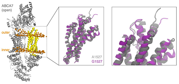
Behold ABCA7. This structure of ABCA7 protein highlights the Gly1527 mutation that increases a person’s risk for AD. [Courtesy of von Maydell et al., Nature, 2025.]
The investigators linked the ABCA7 variants to expression changes in 2,389 genes. These related to cellular stress, apoptosis, synaptic function, DNA repair, and metabolism. Microglia and astrocytes beefed up expression of transcriptional regulators, and microglia also toned down cell stress responses. In oligodendrocytes and their precursor cells, inflammatory gene expression took a hit, as did cellular stress responses.
Neurons also toned down genes related to cellular stress responses. Ditto for synaptic transmission, but they bolstered DNA repair. Excitatory neurons, specifically, carried a distinct transcriptomic fingerprint. They expressed more ABCA7 than did any other cell type. To Tsai, the elevated levels of ABCA7 in neurons was unexpected. “Despite the recent emphasis on glial cells in genetic Alzheimer’s risk, our single-cell data highlighted neurons as key mediators of ABCA7 risk,” she wrote to Alzforum.
Because ABCA7 proved so abundant in excitatory neurons, the researchers focused on 268 genes differentially expressed in these cells, using gene set enrichment analysis to identify perturbed biological pathways. This indicated that these neurons let lipid metabolism slide, while amping up mitochondria and DNA repair and replication. Other pathways were compromised by both up- and downregulated genes, including proteasomal degradation, apoptosis, and synaptic function.
Would the more common ABCA7 risk variants evoke similar responses? Most of these are single nucleotide polymorphisms in the coding region. To address this, von Maydell and colleagues used snRNA-Seq to chart the transcriptomes of excitatory neurons carrying the common p.Ala1527Gly variant, which moderately increases risk for AD. They obtained 360 new cortical tissue samples, of which 133 were from carriers, the remainder, controls. In short, the common, moderate-risk ABCA7 variants evoked the same transcriptome changes as the LoF mutations had.
To see if these snRNA-Seq changes have physiological consequences, the scientists made two isogenic lines of excitatory neurons from iPSC cells and tested their synaptic function. One carried p.Glu5Ofs*3, a frameshift mutation in ABCA7, and the other p.Tyr622*, a nonsense mutation. Each change a single amino acid, glutamic acid and tyrosine respectively, creating a stop codon that truncates the ABCA7 protein, rendering it useless. Per their transcriptomes, both lines were very similar. Pathways related to mitochondria and lipid metabolism mirrored those seen in excitatory neurons in ABACA7 variant postmortem tissue. Both strains of neurons were hyperactive, firing action potentials at lower membrane potentials than healthy neurons. This behavior mimics what goes on in the AD brain, suggesting that ABCA7 mutations cause excitatory neurons to malfunction by compromising lipid metabolism and/or mitochondria.
Next, von Maydell and colleagues homed in on the latter. In p.Glu5Ofs*3 and p.Tyr622* neurons, the powerhouse organelles struggled to maintain their membrane potential and to uncouple electron transport from ATP synthesis. Uncoupling prevents generation of reactive oxygen species, and these cells showed signs of oxidative stress. The mitochondrial transcriptome reflected these functional deficits. Of 1,136 mitochondrial genes, the organelles activated those involved in apoptosis and oxidative phosphorylation and suppressed those controlling metabolism and oxidative detoxification.
Next, the authors found that neutral lipids, phospholipids, sphingolipids, and steroids were off kilter in both induced neuron cultures. Levels of long-chain polyunsaturated triglycerides were up. The p.Glu5Ofs*3 mutation affected phosphatidylcholines (PCs) in different ways. Saturated PCs shot up while highly saturated polyunsaturated fatty acids-containing PCs fell. The findings dovetail with a manuscript posted to bioRxiv on September 7 reporting that ABCA7 variants disrupt lipid profiles, allowing lipid droplets to accumulate in cultured neurons (Nam et al., 2025). Lipid droplets also accumulate in microglia in the aging brain and in AD contexts (Aug 2019 news; Sep 2025 news).
Can restoring lipid homeostasis reverse the transcriptional and functional changes in excitatory neurons? To answer this question, von Maydell and colleagues supplemented p.Glu5Ofs*3 and p.Tyr622* neurons with 5'-diphosphocholine (CDP-choline), which is metabolized in cells to release choline. After two weeks, lipid homeostasis in both ABCA7 neuron lines was back to normal, with unsaturated PCs and neutral lipids at levels seen in isogenic control neurons.
Furthermore, CDP-choline partially restored the transcriptome signature to that of healthy neurons. The mitochondrial membrane potential fell, uncoupling was restored, and the cells showed fewer signs of oxidative stress. Mitochondrial gene expression even looked normal. Action potentials and synaptic activity were back to wild-type patterns.
To Takahisa Kanekiyo, Mayo Clinic, Rochester, Minnesota, that CDP-choline was able to reverse defects caused by ABCA7 loss of function was important. Kanekiyo’s lab also reported that these variants reduced PC and sphingomyelin, another lipid. “It would be also interesting to investigate how sphingolipid metabolism is involved in the mitochondria dysfunction induced by these ABCA7 LOF variants,” he wrote to Alzforum (comment below).
Tsai sees therapeutic potential. “Although CDP-choline is one way to act on these pathways, our work, together with others’, provides a foundation for developing more targeted treatments in the future,” she wrote.—Holly Korthas
References
News Citations
- Newly Identified Microglia Contain Lipid Droplets, Harm Brain
- AD Gene PICALM Hampers Phagocytosis, Promotes Lipid Droplets in Microglia
Paper Citations
- Pericak-Vance MA, Nam Y, DeRosa BA, Ramirez AM, Ayele BA, Whitehead PG, Adams LD, Golightly CG, Starks TD, Laverde-Paz J, Cukier HN, Akinyemi R, Sarfo F, Akpalu A, Cuccaro ML, Williams S, Caban-Holt A, Reitz C, Haines JL, Goldie BS, Rajabli F, Dykxhoorn DM, Young JI, Vance JM. Disrupted Lipid Homeostasis as a Pathogenic Mechanism in ABCA7-Associated Alzheimers Disease Risk. 2025 Sep 07 10.1101/2025.09.03.673792 (version 1) bioRxiv.
External Citations
Further Reading
No Available Further Reading
Primary Papers
- von Maydell D, Wright SE, Pao PC, Staab C, King O, Spitaleri A, Bonner JM, Liu L, Yu CJ, Chiu CC, Leible D, Ni Scannail A, Li M, Boix CA, Mathys H, Leclerc G, Menchaca GS, Welch G, Graziosi A, Leary N, Samaan G, Kellis M, Tsai LH. ABCA7 variants impact phosphatidylcholine and mitochondria in neurons. Nature. 2025 Sep 10; Epub 2025 Sep 10 PubMed.
- Pericak-Vance MA, Nam Y, DeRosa BA, Ramirez AM, Ayele BA, Whitehead PG, Adams LD, Golightly CG, Starks TD, Laverde-Paz J, Cukier HN, Akinyemi R, Sarfo F, Akpalu A, Cuccaro ML, Williams S, Caban-Holt A, Reitz C, Haines JL, Goldie BS, Rajabli F, Dykxhoorn DM, Young JI, Vance JM. Disrupted Lipid Homeostasis as a Pathogenic Mechanism in ABCA7-Associated Alzheimers Disease Risk. 2025 Sep 07 10.1101/2025.09.03.673792 (version 1) bioRxiv.
- Wang Y, Guan X, Chen X, Cai Y, Ma Y, Ma J, Zhang Q, Dai L, Fan X, Bai Y. Choline Supplementation Ameliorates Behavioral Deficits and Alzheimer's Disease-Like Pathology in Transgenic APP/PS1 Mice. Mol Nutr Food Res. 2019 Sep;63(18):e1801407. Epub 2019 Jul 18 PubMed.
- Akgun B, Gulyayev AV, Coker M, Hamilton-Nelson KL, Olalusi O, Adams LD, Akinwande K, Whitehead PG, Diala S, Ogunronbi M, Laux RA, Akpalu A, Starks TD, Sarfo FS, Chavez J, Lwere K, Beecham GW, Akinyemi JO, Obiako R, Ndetei D, Caban-Holt A, Ayele B, Ogrocki P, Griswold AJ, Zaman AF, Mena PR, Martinez IM, Blanton SH, Okubadejo N, Damasceno A, Kalaria R, Cuccaro ML, McInerney KF, African Dementia Consortium (AfDC), Alzheimer's Disease Sequencing Project (ADSP), Bush WS, Kunkle BW, Ogunniyi A, Vance JM, Akinyemi RO, Haines JL, Reitz C, Byrd GS, Rajabli F, Pericak-Vance MA. ABCA7 deletion lowers age at onset of Alzheimer's disease and interacts with APOE ε4 synergistically in African-ancestry populations. Alzheimers Dement. 2025 Aug;21(8):e70583. PubMed.
Annotate
To make an annotation you must Login or Register.
Comments
Mayo Clinic
Maydell et al. have demonstrated that ABCA7 loss-of-function (LOF) variants disrupt phosphatidyl choline metabolism and mitochondrial homeostasis in human neurons. Using single-nucleus RNA-Seq in postmortem AD brains with ABCA7 LOF variants, and iPSC-derived neuronal models, the authors provide strong evidence that ABCA7 dysfunction in neurons contributes to AD risk through metabolic and bioenergetic pathways, consistent with our previous findings through ABCA7 knockout iPSC models.
Importantly, CDP-choline supplementation could rescue these defects caused by ABCA7 LOF. CDP-choline could also ameliorate APOE4-related phenotypes, highlighting its therapeutic potential for AD. This compound also increased sphingomyelin in the ABCA7 LOF iPSC neurons in the study. Thus, it would be interesting to investigate how sphingolipid metabolism is involved in the mitochondria dysfunction induced by these ABCA7 variants. Indeed, in our study ABCA7 deficiency reduced PC and SM in iPSC-derived cortical organoids. Future studies exploring the lipid metabolic pathway affected by other AD risk genes, as well as ABCA7, may provide clues to develop novel lipid-targeting therapies for AD.
Make a Comment
To make a comment you must login or register.