
Could Monocyte-Derived Microglia Cause Some Cases of Parkinsonism?
Quick Links
Microglia in the brain’s parenchyma arise from macrophage progenitors in the embryonic yolk sac (Ginhoux et al., 2010), and are thought to self-renew locally during a mammal’s lifespan, without macrophages from the hematopoietic system adding to their numbers.
- Certain brain regions of healthy old mice and humans harbor monocyte-derived microglia.
- The transcriptome of some monocyte-derived microglia was identical to that of yolk sac-derived microglia.
- Monocyte-derived microglia bearing a DNMT3A gene variant caused motor deficits resembling parkinsonism.
However, an international collaboration led by Jung-Seok Kim and Steffen Jung from the Weizmann Institute of Science in Rehovot, Israel, reported that as mice age, monocyte-derived microglia accumulate in their brains, where they express the same genes as yolk sac-derived microglia. In a mouse transplantation model, mutated monocytes even caused motor deficiencies that resemble Parkinson’s disease in mice.
“My take is that in biology, nothing is exclusive. There are always exceptions,” said Jung, senior author the paper, published in Cell Reports on May 27.
“The origin of microglia seemed to be set in stone after the publication of the milestone paper from Florent Ginhoux and Miriam Merad,” writes Amanda Sierra of the Achucarro Basque Center for Neuroscience in Leioa, Spain. “Now, the established paradigm is questioned again,” writes Sierra (comment below).
In the brain’s parenchyma, scientists generally do not see monocyte-derived macrophages become yolk sac-derived microglia. In various experimental settings, monocyte-derived macrophages have occasionally survived in the brain parenchyma, but they tend to disappear and never develop the exact identity of yolk sac-derived microglia (Ajami et al., 2007; Ajami et al., 2011; Shemar et al., 2018; Bennett et al., 2018; Cronk et al., 2018).
To track blood-derived macrophages in the brain, the scientists used Ms4a3Cre:R26-TdTomato mice, whose parenchymal macrophages express a fluorescent label whenever they express Ms4a3, a marker of granulocyte monocyte progenitor-derived monocytes. Flow cytometric analysis of their brains at different ages showed a monocyte-derived macrophage signal that grew with time. Using young and old double-reporter mice that enable distinction between monocyte-derived and yolk sac-derived macrophages, the scientists saw that monocyte-derived macrophages accumulate in the brain parenchyma as mice age.

Present in Parenchyma. In double reporter mice, microglia-like macrophages derived from monocytes (red) co-exist with yolk sac-derived microglia (green).
Using single-cell transcriptome analysis, the scientists confirmed that about 80 percent of the monocyte-derived microglia are indistinguishable from the yolk sac-derived microglia in the double reporter mice brain parenchyma. Some of these macrophages even started looking like microglia morphologically.
Monocyte-derived microglia arise from hematopoietic stem cells, which divide throughout a mammal’s lifetime to generate monocytes. Over time, these stem cells accrue mutations, for example in DNM3TA, a gene encoding a DNA methylation enzyme. Jung and colleagues used such mutations as barcodes to identify mutated monocyte-derived macrophages from among the macrophages in aged mouse brain tissue.
To learn whether this mutation might affect function, the team set up an experiment with two groups of mice. One group were engrafted with bone marrow from mice that had monocytes with this DNM3TA mutation, while the control group had wild-type microglia and wild-type monocyte-derived microglia.
The scientists saw that the mutated monocyte-derived microglia did not settle everywhere in the aging mouse brain. None were to be found in the frontal cortex, but pockets of them were in the midbrain and brainstem (image below).
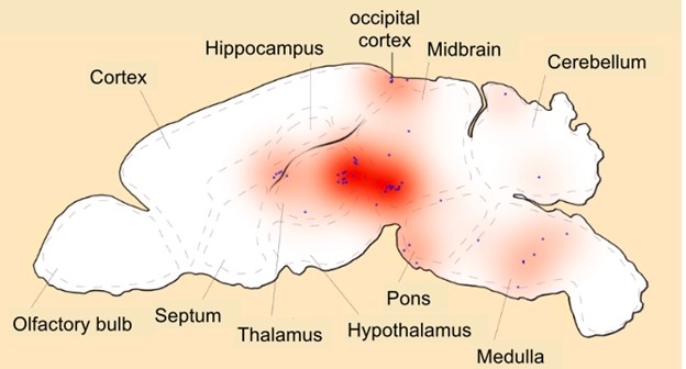
Spotty Settlements. Most monocyte-derived macrophages were in midbrain and medulla, regions affected in parkinsonian diseases.
Can monocyte-derived microglia affect the function of the aging mouse brain? Testing the mice’s behavior, Jung and colleagues measured a deficit in the cat-walk test, in which mice walk over a glass plate while their gait is being recorded. Analysis of this data, collected two, three, and four months after engraftment, showed that mice whose monocyte-derived microglia did not carry the hematopoiesis variant walked evenly at all time points. After four months, the mice that did harbor the mutant monocyte-derived microglia slowed down and fell out of rhythm. This signaled motor deficits, hinting at dysfunction of dopaminergic circuits. Subsequent analysis of 7-month old mouse brain tissue indicated a loss of dopaminergic neurons in the niagrostriatal region affected in Parkinson’s disease.
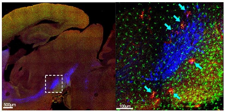
Parkinsonian Pathology? In 7-month-old double reporter mice, monocyte-derived microglia (red) settle in the substantia nigra near neurons that express tyrosine hydroxylase (blue).
Curiously, the LRRK2 gene, mutations of which are a frequent genetic cause of Parkinson’s, is strongly expressed in macrophages.
“The striking finding that these cells can carry somatic mutations associated with clonal hematopoiesis—and potentially contribute to brain pathology—opens exciting new avenues for research,” writes Marco Colonna from Washington University (comment below).
“The paper opens many questions about the mechanisms through which monocyte-derived cells get to reside in the brain. It is not clear whether there is continuous infiltration of monocytes, local proliferation of early infiltrates, or both,” writes Sierra.
See also related story, story.—Andrea Tamayo
Andrea Tamayo is a freelance writer living in Brooklyn, New York.
References
News Citations
- Replacing Rogue Microglia Treats Monogenic Brain Disease Krabbe
- Circulating Monocytes Replace Microglia, Border-Associated Macrophages
Paper Citations
- Ginhoux F, Greter M, Leboeuf M, Nandi S, See P, Gokhan S, Mehler MF, Conway SJ, Ng LG, Stanley ER, Samokhvalov IM, Merad M. Fate mapping analysis reveals that adult microglia derive from primitive macrophages. Science. 2010 Nov 5;330(6005):841-5. PubMed.
- Ajami B, Bennett JL, Krieger C, Tetzlaff W, Rossi FM. Local self-renewal can sustain CNS microglia maintenance and function throughout adult life. Nat Neurosci. 2007 Dec;10(12):1538-43. PubMed.
- Ajami B, Bennett JL, Krieger C, McNagny KM, Rossi FM. Infiltrating monocytes trigger EAE progression, but do not contribute to the resident microglia pool. Nat Neurosci. 2011 Sep;14(9):1142-9. PubMed.
- Shemer A, Grozovski J, Tay TL, Tao J, Volaski A, Süß P, Ardura-Fabregat A, Gross-Vered M, Kim JS, David E, Chappell-Maor L, Thielecke L, Glass CK, Cornils K, Prinz M, Jung S. Engrafted parenchymal brain macrophages differ from microglia in transcriptome, chromatin landscape and response to challenge. Nat Commun. 2018 Dec 6;9(1):5206. PubMed.
- Bennett FC, Bennett ML, Yaqoob F, Mulinyawe SB, Grant GA, Hayden Gephart M, Plowey ED, Barres BA. A Combination of Ontogeny and CNS Environment Establishes Microglial Identity. Neuron. 2018 Jun 27;98(6):1170-1183.e8. Epub 2018 May 31 PubMed.
- Cronk JC, Filiano AJ, Louveau A, Marin I, Marsh R, Ji E, Goldman DH, Smirnov I, Geraci N, Acton S, Overall CC, Kipnis J. Peripherally derived macrophages can engraft the brain independent of irradiation and maintain an identity distinct from microglia. J Exp Med. 2018 Jun 4;215(6):1627-1647. Epub 2018 Apr 11 PubMed.
Further Reading
No Available Further Reading
Primary Papers
- Kim JS, Trzebanski S, Shin SH, Schori L, Frumer Friedman GR, Ilani NC, Kadam A, Vicario R, Aust O, Bugaeva P, Piatek S, Ismajli LK, Hoffmann CJ, Scheller M, Boura-Halfon S, Kaushansky N, Golani O, Solomon A, Liu Z, Amann L, Böhm-Sturm P, Koch SP, Wenger N, Netherlands Brain Bank, Ginhoux F, Prinz M, Avraham R, Harms C, Geissmann F, Müller-Tidow C, Uderhardt S, Milenkovic I, Shlush L, Jung S. Clonal hematopoiesis-associated motoric deficits caused by monocyte-derived microglia accumulating in aging mice. Cell Rep. 2025 May 27;44(5):115609. Epub 2025 Apr 24 PubMed.
Annotate
To make an annotation you must Login or Register.
Comments
Achucarro Basque Center for Neuroscience
The origins of microglia as macrophages resident in the brain parenchyma have been the subject of a heated controversy since their discovery by Rio-Hortega in 1919, when researchers debated whether they were mesodermic, as Rio-Hortega proposed, or ectodermic. For most of the 20th century, scientists still debated whether microglia came from circulating monocytes, as suggested by irradiation followed by bone marrow transplantations. Then, a breakthrough paper from Bahareh Ajami and Fabio Rossi at the University of British Columbia used parabiosis to show that when the blood-brain barrier was undisturbed (as opposed to what occurs during irradiation), monocyte-derived cells were not found in the parenchyma. The origin of microglia seemed to be set in stone after the publication of the milestone paper from Florent Ginhoux and Miriam Merad at the Mount Sinai School of Medicine, who used lineage tracing to identify microglial precursors in the yolk sac. Now, the established paradigm is questioned again by Jung-Seok Kim and Steffen Jung at the Weizmann Institute by showing that, after all, monocyte-derived microglia cells exist in the brain parenchyma and accumulate with age.
Kim et al.’s most salient finding is the identification of monocyte-derived microglia residing in the brain parenchyma, using a sophisticated lineage tracing strategy based on the Ms4a3 promoter, which is specifically expressed in monocytes derived from bone marrow granulocyte-monocyte progenitors. The authors then used scRNA-sequencing to demonstrate that the incoming cells expressed the transcriptional signature of microglia. While these cells were rare in young brains, they accumulated in older brains in regions such as the hypothalamus and the nigrostriatal system, although surprisingly, the cortex was spared.
These results prompted the authors to test age-related phenotypes caused by monocyte-derived microglia, and they focused on somatic mutations, which are known to accumulate during aging in hematopoietic cells as a result of clonal hematopoiesis. These mutations frequently affect genes related to epigenetic regulation, such as DNMT3A. Introducing a common human DNMT3A variant in mouse monocytes led to a Parkinsonian syndrome, with reduced numbers of dopaminergic neurons in the substantia nigra and motor deficits, an effect presumably mediated by microglia-derived monocytes, although the authors could not disregard an effect of circulating monocytes or lymphocytes carrying the mutant DNMT3A. Finally, the authors indirectly showed that human brains could also harbor monocyte-derived microglia by comparing the frequency of clonal hematopoiesis variant alleles in blood and in Pu.1 cells from the brain. As Pu.1 labels all brain macrophages (microglia from either yolk sac or from monocytes, and border-associated macrophages), a “comparable” frequency of clonal hematopoiesis variant alleles was interpreted as evidence of the presence of monocyte-derived cells residing in the human brain.
Incidental observations from us and others suggested that in some disease models, infiltrating monocytes can turn into microglia. For example, in a stroke model using CCR2-RFP/CX3CR1-EGFP mice, in which blood monocytes express the red reporter and parenchymal microglia the green reporter, we observed yellow cells over time, suggesting that monocytes acquired microglia features (Beccari et al., 2023). Now Jung and colleagues have demonstrated the presence of these cells in unperturbed brains, suggesting that the brain contains a mosaic of microglia of different origins.
The paper opens many questions about the mechanisms through which monocyte-derived cells get to reside in the brain. It is not clear whether there is continuous infiltration of monocytes, local proliferation of early infiltrates, or both. It is also unclear why monocytes home to some particular brain regions—the authors speculate that there could be differences in the blood-brain barrier permeability, but it could also be interesting to determine whether local neuronal activity is related to the infiltration and/or expansion of these cells. Another aspect that remains to be determined is the interaction of the invading monocytes with the resident cells, because yolk sac-derived microglia have a tessellated distribution that occupies the whole space, and it is only when their niche is empty that other cells (repopulating cells, transplanted cells) get to settle down. While the authors have shown that monocyte-derived microglia acquire a transcriptional profile similar to that of yolk-derived cells, it remains to be tested whether they are functionally equivalent, in terms of process motility and brain surveillance, inflammatory responses, and phagocytosis of different types of cargo. Finally, a more direct method of assessment of human monocyte-derived microglia is necessary to confirm their presence and implication in the physiology and/or pathology of the human brain.
References:
Beccari S, Sierra-Torre V, Valero J, Pereira-Iglesias M, García-Zaballa M, Soria FN, De Las Heras-Garcia L, Carretero-Guillen A, Capetillo-Zarate E, Domercq M, Huguet PR, Ramonet D, Osman A, Han W, Dominguez C, Faust TE, Touzani O, Pampliega O, Boya P, Schafer D, Mariño G, Canet-Soulas E, Blomgren K, Plaza-Zabala A, Sierra A. Microglial phagocytosis dysfunction in stroke is driven by energy depletion and induction of autophagy. Autophagy. 2023 Jul;19(7):1952-1981. Epub 2023 Jan 20 PubMed.
Washington University School of Medicine
Kim et al. demonstrate that monocyte-derived microglia (MoMg) accumulate in the brains of aging mice, particularly in the nigrostriatum and medulla. These MoMg adopt the morphology and gene expression profiles characteristic of microglia despite their monocytic origin, including the expression of Sall1. However, unlike yolk sac-derived embryonic microglia, MoMg can undergo clonal hematopoiesis due to their hematopoietic stem cell origin. In this context, MoMg could be pathogenic. To support this hypothesis, they authors use a chimeric transfer model to show that MoMg carrying the DNMT3AR882H mutation, a key variant in human clonal hematopoiesis, lead to motor impairments resembling atypical Parkinsonian disease.
While aging may lead to partial replacement of microglia by bone marrow–derived MoMg, the striking finding that these cells can carry somatic mutations associated with clonal hematopoiesis—and potentially contribute to brain pathology—opens exciting new avenues for research.
Make a Comment
To make a comment you must login or register.