
Replacing Rogue Microglia Treats Monogenic Brain Disease Krabbe
Quick Links
Replacing rogue microglia with healthy ones can ameliorate, in a mouse model, a childhood neurodegenerative disease called Globoid cell leukodystrophy (GLD), aka Krabbe disease. The approach even extended survival, according to researchers led by Chris Bennett at the University of Pennsylvania in Philadelphia. On May 13 in Immunity, the team reported that the replacement reduces the number of toxic giant cells, called globoid cells, that build up in the disease.
- Interferon at root of growing microglial mayhem in Krabbe mouse model.
- Study characterizes molecular signature of pathological macrophages in mice and humans.
- Transplanted monocyte-derived microglia lessen pathogenesis in Krabbe mouse.
“These findings strongly support the therapeutic potential for microglia replacement strategies for GLD and offer valuable insights into the mechanisms underlying GLD-related microglia dysfunction,” Jean Paul Chadarevian of the University of California, Irvine, wrote to Alzforum.
Why study this rare disease? The scientists note four reasons. It is caused by loss-of-function mutations in the gene for the enzyme galactocerebrosidase (Galc). Already in early childhood, these mutations lead to myelin damage and cellular lipid accumulations somewhat similar to changes seen much later in life in neurodegenerative diseases such as Alzheimer’s and Parkinson’s. In Krabbe, macrophages lacking Galc accumulate to form toxic globoid cells, after which GCD was named. Twitcher, a mouse model with a Galc missense mutation, develops a tremor 20 days after birth, and globoid cells in the brain as the disease progresses. In children, GCD is being treated with bone marrow transplants, enabling the scientists to compare that treatment to more targeted cell therapies, e.g., monocyte transplant.
To start, first author William Aisenberg and colleagues sought to understand how the macrophages in the twitcher mouse change over time. To do so, they bulk-sequenced the RNA of the macrophages in twitcher and wild-type mice before, during, and after symptom onset. Starting at day 15, interferon-stimulated genes became differentially expressed in twitcher macrophages. Over time, gene expression in brain macrophages turned increasingly abnormal, suggesting that macrophage dysfunction may begin with interferons and morph through a continuum of reactive states. The scientists validated this change sequence in macrophage gene expression signature by way of in situ RNA hybridization in brain tissue of twitcher and wild-type mice.
Next, Aisenberg and colleagues characterized globoid cells, a descriptive name for the microscopically distinctive pathogenic brain immune cells that start to show up when young children with Krabbe develop symptoms. In 1916, Danish neurologist Knud Krabbe first described these cells as “gigantic polynuclear glia-cells with big fibres and degenerated nuclei.”
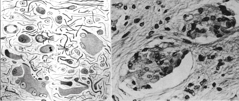
Generations of Globoids. On the left, globoid cells as drawn by neurologist Knud Krabbe. On the right, a micrograph of these cells from 1973. [Courtesy of Alastair Compston, 2013, and Jacob et al., 1973.]
The standard tool pathologists use to recognize these cells is a Periodic Acid-Schiff (PAS) stain, but the cells’ molecular identity remained undefined. Now there are better tools to characterize them, said Aisenberg, a postdoc in Bennett’s lab. Again using RNA in situ hybridization, the team probed for four genes expressed by human and mouse globoid cells: Lgals3, Ms4a7, Gpnmb, and Spp1. This gave them their first molecular signature and definition.
Some of the genes identified are associated with Alzheimer’s disease. “Macrophage or microglial dysfunction in AD might be due to dysfunctional lipid metabolism and homeostasis,” Aisenberg said. α-Synuclein, a protein involved in Parkinson’s and Lewy body dementia, has also been found to aggregate and contribute to Krabbe disease progression (Hatton et al., 2022).
“The first part of our paper is characterizing the role of macrophages in a disease state. The second half asks, ‘How does delivering therapeutic macrophages change or not change that?’” Aisenberg told Alzforum.
To address this, the scientists first depleted twitcher mouse microglia via tamoxifen and followed that by injecting GFP-labeled monocytes into the mouse brains. This repopulated the brains with macrophages that express Galc and glow under the microscope.
With in situ hybridization, the team probed for three genes globoid cells express––Ms4a7, Lgals3, and Fn1 in wild-type, twitcher, and twitcher mice treated with a monocyte transplant. They found that the latter didn’t express Lgals3 and Fn1; the only one they did express, Ms4a7, is a marker of monocyte-origin macrophages. More broadly, treated twitcher mice matched the wild-type, i.e., they didn’t develop the toxic giant globoid cells, nor the same disease state as untreated mice.

Goodbye Globoid. Globoid cell clusters in twitcher mice express Ms4a7, Lgals3, and Fn1 (second row). Similar to wild-type parenchymal macrophages (top row), macrophages implanted into twitcher brains (third row) don’t exhibit Lgals3 (yellow) or Fn1 colocalized with IBA1, a marker of macrophages and microglia (pink), but they do show the monocyte marker Ms4a7 (blue). [Courtesy of Aisenberg et al., Immunity, 2025.]
PAS staining confirmed that the Krabbe mice with transplanted monocytes had no globoid cells in their brainstems and cerebelli, which are affected in Krabbe patients. The twitcher brain environment appeared not to affect the transplanted cells. This surprised Aisenberg because tissue macrophages are sensitive to their environments. Overall, the scientists found that twitcher mice with the transplanted monocytes lived for 80 days, as compared to 40 days for those without the transplant. Along the way, treated mice accumulated less toxic lipid, retained more myelinated axons, and had reduced astrogliosis.
“These findings hold significant translational potential,” Costantino Iadecola of Cornell Weill Medical College in New York City wrote to Alzforum. He was not involved in this work.
Many questions remain, Aisenberg said, for example whether globoid cells are helpful or harmful. One hypothesis holds that they are big, reactive macrophages that are driving pathogenesis; the other, that they become engorged as they sequester toxic lipids. If the latter, removing them could worsen disease. “Now that we have this molecular definition of a globoid cell, we can use genetic tools to disrupt their formation and function and ask: What are they doing in the disease?” questions Aisenberg.
See also related story, story.—Andrea Tamayo
Andrea Tamayo is a freelance writer living in Brooklyn, New York.
References
News Citations
- Circulating Monocytes Replace Microglia, Border-Associated Macrophages
- Could Monocyte-Derived Microglia Cause Some Cases of Parkinsonism?
Paper Citations
- Compston A. A new familial infantile form of diffuse brain-sclerosis. Brain. 2013 Sep;136(Pt 9):2649-51. PubMed.
- Jacob JC, Kutty KM, Islam M, Dominic RG, Dawson G. Krabbe's disease: globoid cell leukodystrophy. Can Med Assoc J. 1973 Jun 2;108(11):1398-400. PubMed.
- Hatton C, Ghanem SS, Koss DJ, Abdi IY, Gibbons E, Guerreiro R, Bras J, International DLB Genetics Consortium, Walker L, Gelpi E, Heywood W, Outeiro TF, Attems J, McFarland R, Forsyth R, El-Agnaf OM, Erskine D. Prion-like α-synuclein pathology in the brain of infants with Krabbe disease. Brain. 2022 May 24;145(4):1257-1263. PubMed.
Further Reading
Primary Papers
- Aisenberg WH, O'Brien CA, Sangster M, Yaqoob F, Zhang Y, Temsamrit B, Thom S, Gosse L, Chaluvadi S, Elfayomi B, Lee G, Polam V, Levitt EM, Liu G, Lombroso SI, Nemec KM, Clowry G, Nieves C, Rawat P, Church E, Martinez D, Shoffler C, Kancheva D, Petucci C, Taylor D, Kofler J, Erskine D, Movahedi K, Bennett ML, Bennett FC. Direct microglia replacement reveals pathologic and therapeutic contributions of brain macrophages to a monogenic neurological disease. Immunity. 2025 May 13;58(5):1254-1268.e9. Epub 2025 Apr 30 PubMed.
Annotate
To make an annotation you must Login or Register.
Comments
University of California, Irvine
The Aisenberg et al. and Bastos et al. studies provide valuable new insights into the complex roles of resident and infiltrating macrophages in the brain, and they highlight the potential of microglia replacement therapies for the treatment of neurological diseases.
Bastos et al. demonstrate that peripherally derived monocytes can efficiently engraft and replace both resident and border-associated macrophages following microglia depletion. A key finding demonstrates that fetal liver monocytes, with their distinct epigenetic landscape, can acquire Sall1-positive expression, a key marker of microglial identity not observed by infiltrating bone-marrow derived monocytes. Leveraging their transcriptomic profiling of monocyte-derived microglia, this study also identifies infiltrating monocytes in postmortem brain tissue from Alzheimer’s disease (AD) patients surrounding amyloid plaques. These findings underscore the relevance of monocyte recruitment in AD and that macrophage ontogeny profoundly influences their fate and functional potential within the brain.
Aisenberg et al. present a comprehensive study on the role of brain macrophages in Globoid Cell Leukodystrophy (GLD), also known as Krabbe disease. Using the twitcher mouse model, the authors generate a single-cell transcriptomic atlas of Twi macrophages, revealing that the shift from normal microglial maturation to a progressive state of dysfunction is marked by an early induction of interferon-stimulated genes.
Interestingly, this study demonstrates direct monocyte transplantation into the brain of microglia-depleted Twi-neonates normalizes a distinct molecular signature of globoid cells, extends survival, improves myelin integrity, and normalizes other GLD-hallmark pathologies. These findings strongly support the therapeutic potential for microglia replacement strategies for GLD and offer valuable insights into the mechanisms underlying GLD-related microglia dysfunction.
Together, these two studies further our understanding of brain macrophage biology and support the development of microglia-targeting therapies for a variety of neurological disorders. Bastos et al. profile alternative microglia donors, while Aisenberg et al. demonstrate its potential efficacy in a disease-specific context. Together, they raise exciting possibilities for the development of microglia replacement strategies for human disease.
References:
Aisenberg WH, O'Brien CA, Sangster M, Yaqoob F, Zhang Y, Temsamrit B, Thom S, Gosse L, Chaluvadi S, Elfayomi B, Lee G, Polam V, Levitt EM, Liu G, Lombroso SI, Nemec KM, Clowry G, Nieves C, Rawat P, Church E, Martinez D, Shoffler C, Kancheva D, Petucci C, Taylor D, Kofler J, Erskine D, Movahedi K, Bennett ML, Bennett FC. Direct microglia replacement reveals pathologic and therapeutic contributions of brain macrophages to a monogenic neurological disease. Immunity. 2025 May 13;58(5):1254-1268.e9. Epub 2025 Apr 30 PubMed.
Bastos J, O'Brien C, Vara-Pérez M, Mampay M, van Olst L, Barry-Carroll L, Kancheva D, Leduc S, Lievens AL, Ali L, Vlasov V, Meysman L, Shakeri H, Roelandt R, Van Hove H, De Vlaminck K, Scheyltjens I, Yaqoob F, Lombroso SI, Breugelmans M, Faron G, Gomez-Nicola D, Gate D, Bennett FC, Movahedi K. Monocytes can efficiently replace all brain macrophages and fetal liver monocytes can generate bona fide SALL1+ microglia. Immunity. 2025 May 13;58(5):1269-1288.e12. Epub 2025 Apr 30 PubMed.
Make a Comment
To make a comment you must login or register.