Dipeptide Repeats May Hobble Ribosomes in C9ORF72-FTD Patient Brain
Quick Links
How do hexanucleotide expansions in the C9ORF72 gene cause amyotrophic lateral sclerosis and frontotemporal dementia? According to a study published May 16 in Life Science Alliance, a new open-access journal run by EMBO Press, Rockefeller Press, and Cold Spring Harbor Laboratory Press, the dipeptide repeats translated from the expansions glom onto ribosomes, hindering protein translation. Researchers led by Dieter Edbauer, German Center for Neurodegenerative Diseases, Munich, screened for proteins that interact with the dipeptide repeats and pulled out numerous potential mediators of toxicity in cultured neurons. However, only ribosomal proteins stood out in postmortem brain samples from C9-FTD patients. The findings suggest that a gradual erosion of protein synthesis could mark the long prodromal phase of these neurodegenerative diseases. They also caution against drawing conclusions about toxicity based solely on screens performed in cultured cells, Edbauer said.
- Poly-GR/PR dipeptide repeats interact with stress granules, RNA-binding, ribosomal, and many proteins in cell culture.
- Repeats predominantly bound up with ribosomes in patient brain.
- Poly-GR/PR may hurt neurons by hindering protein translation.
Dipeptide repeats (DPRs) congregate in cytoplasmic inclusions in neurons from ALS/FTD patients. The polypeptides arise from translation in six reading frames, yielding five flavors of peptide: glycine-alanine (GA), glycine-arginine (GR), proline-arginine (PR), proline-alanine (PA), and glycine-proline (GP). Of these, the arginine-containing varieties—poly-GR and poly-PR—are thought to be most toxic. Researchers have used screens to uncover potential modifiers of polyGR/PR toxicity, pointing to proteins known to mingle in membraneless organelles, including the nucleolus and stress granules (Oct 2016 news; Boeynaems et al., 2016; and Mar 2018 news). However, do these interactions take place in the brain? And are they toxic there?
Co-first authors Hannelore Hartmann and Daniel Hornburg set out to find proteins that interact with DPRs, and then to confirm those interactions in patients' brains. They used lentiviral vectors to overexpress fluorescently tagged DPRs (either 149 repeats of GR, or 175 repeats of PR) in primary rat cortical neurons. Right off the bat, they found dramatic differences in localization. Poly-PR resided predominantly in the nucleolus, a membrane-less organelle where ribosomes are made, while poly-GR was spread diffusely throughout the cytoplasm with only a minor presence in the nucleolus. Using immunoprecipitation followed by mass spectrometry, the researchers fished out 89 proteins that interacted with poly-PR, and 104 that associated with poly-GR. Thirty-nine overlapped. Despite their distinct cellular locales, both DPRs interacted with RNA-binding proteins, as well as numerous components of ribosomes, the nucleolus, and stress granules. The researchers pulled out a similar list of polyGR/PR partners when performing screens in HEK293 cells.
The list of polyGR/PR interaction partners came as no surprise, as it corroborated those of previous studies. Reviewers’ comments—available on the journal’s website—cited this redundancy as detracting from the novelty of the paper. However, Edbauer told Alzforum that his main goal was to determine which of these binding partners hooked up with DPRs in the brain.
Before doing that, the scientists tried to validate six top hits by overexpressing fluorescently tagged versions, and DPRs, in neurons and HEK293 cells. Nucleolar proteins NPM1 and NOP56 co-localized with polyGR/PR in the nucleolus, and appeared to draw in the normally cytoplasmic poly-GR. RNA-binding proteins STAU1 and STAU2 or YBX1 promoted cytoplasmic clustering of DPRs in organelles expressing stress granule markers. The 40S ribosomal protein RPS6 showed a modest association between it and polyGR/PR clusters. These findings suggested that in cultured cells, DPR interactors hailing from different organelles associated with the poly-dipeptides, with the nucleolus and stress granules being prime sites for DPR toxicity.
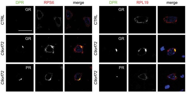
Alas, patient cells told a different story. In postmortem brain samples from two C9-FTD patients, the researchers found nary a single instance of DPR inclusions that co-localized with stress granule markers. They did find inclusions containing STAU2. However, Edbauer told Alzforum that while this RNA-binding protein typically inhabits stress granules, it does not alone define the organelle. Furthermore, the researchers found no accumulation of either DPR in the nucleolus of patient cells. Instead, they saw an abundance of ribosomal proteins intermixed with DPR cytoplasmic inclusions.

Ribosomal Rendezvous. The ribosomal protein RPS6 (red) co-localized with DPRs (yellow, merge) in brain samples from C9-FTD patients (bottom two panels), but not control (top). [Courtesy of Hartmann et al., 2018.]
To define this ribosomal association, the researchers returned to cell culture. Transducing primary neurons with genes for either of the DPRs reduced levels of ribosomal proteins and protein synthesis. These effects were dramatic in cells expressing poly-PR, but subtle in those expressing poly-GR. Poly-PR expression was also far more toxic to neurons than poly-GR, and only poly-PR went into the nucleolus.
Curiously, when the researchers truncated poly-GR from 149 repeats to 53, it behaved more like the 175-mer polyPR, moving into the nucleolus, knocking down ribosomal biogenesis and protein translation, and ultimately killing neurons. Edbauer does not know why shorter poly-GRs occupy the nucleolus. However, the findings suggest that nucleolar localization correlates with cellular toxicity and inhibition of ribosome assembly and thus, protein translation … at least in cultured cells. Back in the patient brain, the researchers observed neither DPR accumulating in the nucleolus, and instead just noticed them interacting with ribosomal proteins in the cytoplasm.
The researchers propose that the DPRs exert toxicity through distinct mechanisms in cellular-overexpression models and in the human brain. In cultured cells, overexpressed DPRs infiltrate the nucleolus, where they interfere with ribosome biogenesis and profoundly suppress protein translation. In the brain, the dipeptides primarily accumulate in the cytoplasm, where they bind to fully assembled ribosomes, interfering with protein translation in a more subtle way. These findings could explain why the DPRs expressed in cultured cells appear to be far more toxic than those in the human brain, which arise long before disease symptoms manifest, Edbauer said. He also said that most researchers use a short version of poly-GR, which more easily enters the nucleolus. Most patients have far longer repeat expansions.
The researchers acknowledged that identifying this interaction in patient cells is but a start, and more work remains to determine if ribosomal interactions are how DPRs are toxic in human disease.
How the polyGR/PR mechanisms intersect with other pathologies is also of interest. Notably, DPRs may have company at the ribosomes, because a previous study reported that TDP-43 also latches onto the protein factories, dampening translation (Feb 2017 news). Furthermore, Edbauer and other researchers previously reported that the DPR poly-GA interferes with the proteasome, suggesting that together, DPRs rattle two pillars of proteostasis in the cell (Feb 2018 news). Edbauer likened the polyGR/PR dipeptides to the role of Aβ in AD. “They are only mildly toxic, so the brain can cope with them for a long time, but eventually the system crashes.”—Jessica Shugart
References
News Citations
- ALS Research ‘Gels’ as Studies Tie Disparate Genetic Factors Together
- CRISPR Screen Pulls Down Fresh Targets for C9ORF72 ALS
- In New Role for TDP-43, Scientists Say it Controls Protein Synthesis
- Are ALS Dipeptide Repeat Ribbons Entangling Proteasomes?
Paper Citations
- Boeynaems S, Bogaert E, Michiels E, Gijselinck I, Sieben A, Jovičić A, De Baets G, Scheveneels W, Steyaert J, Cuijt I, Verstrepen KJ, Callaerts P, Rousseau F, Schymkowitz J, Cruts M, Van Broeckhoven C, Van Damme P, Gitler AD, Robberecht W, Van Den Bosch L. Drosophila screen connects nuclear transport genes to DPR pathology in c9ALS/FTD. Sci Rep. 2016 Feb 12;6:20877. PubMed.
Further Reading
Primary Papers
- Hartmann H, Hornburg D, Czuppa M, Bader J, Michaelsen M, Farny D, Arzberger T, Mann M, Meissner F, Edbauer D. Proteomics and C9orf72 neuropathology identify ribosomes as poly-GR/PR interactors driving toxicity. Life Science Alliance. 16 May 2018. DOI: 10.26508/lsa.201800070
Annotate
To make an annotation you must Login or Register.

Comments
No Available Comments
Make a Comment
To make a comment you must login or register.