
Lipid Droplets: Gatekeepers of Microglial Activation, for Better or Worse
Quick Links
When microglia wind up with a belly full of lipid droplets, it’s a sign that the cells are rallying, perhaps in vain, to a neurodegenerative threat. But according to a study published July 10 in Cell Reports, these fatty deposits are more than bellwethers; rather, they play an essential role in many aspects of microglial activation. Scientists led by Priyanka Narayan of the National Institutes of Health, Bethesda, Maryland, report that whether microglia are riled by an external stimulus, such as lipopolysaccharide, or via internal turmoil, as brought about by expressing ApoE4, the metabolism of triglycerides, carried in lipid droplets, was necessary for the cells to mount a transcriptional response. Blocking triglyceride biosynthesis prevented microglia from churning out cytokines, ramping up phagocytic capacity, and rapidly reorienting their membrane processes. This was true whether microglia were responding to LPS, or festering in a chronic, dysfunctional state thanks to ApoE4.
- If they cannot make triglycerides, microglia do not react to lipopolysaccharide.
- They will not move, release cytokines, or phagocytose debris.
- Blocking fat biosynthesis weakened inflammatory responses of ApoE4 microglia.
“These findings give further support to microglial triglyceride lipid droplet accumulation as an important new drug target in AD, a target strongly supported by human genetics but for which there is no therapeutic,” commented Michael Haney and Sydney Mason of the University of Pennsylvania, Philadelphia.
Both neuropathological and genetic findings place lipid metabolism at the heart of AD. Along with plaques and tangles, lipid granules within glia were among the hallmark pathologies described by Alois Alzheimer in his famed 1907 case report (Alzheimer, 1907). Later, ApoE4, the prime lipid carrier within the brain, was pegged as the strongest genetic AD risk factor, and other genes involved in lipid homeostasis were subsequently tied to the disease via GWAS. Recent studies have taken a fresh look at the role of lipid droplets in AD, reporting that microglia are crowded with them in AD brains, and that the fat-laden cells are dysfunctional and even harmful to neurons (Aug 2019 news; Mar 2021 news; Feb 2025 news).
What role do these lipid storage depots play in microglial activation? First author Roxan Stephenson and colleagues addressed this using, for the most part, iPSC-derived microglia. For a robust model of activation, the scientists stimulated ApoE3/E3 microglia with LPS. As expected, this inflammatory sledgehammer provoked the cells to pump out pro-inflammatory chemokines and cytokines, change shape, and gobble up numerous types of debris, including Aβ. The cells also accumulated lipid droplets and ramped up not only immune signaling genes, but those involved in triglyceride biosynthesis.
To find out how this uptick in lipid production relates to activation, the scientists treated microglia with inhibitors of diacyl-glycerol acid transferases 1 and 2 (DGAT1/2). These enzymes control the rate-limiting step in triglyceride biosynthesis. The inhibitors reduced basal levels of lipid droplets within microglia, and prevented their accumulation in response to LPS (image below).
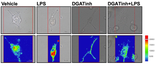
Drowning in Droplets. According to Raman imaging microscopy, LPS provoked accumulation of lipid droplets (red) in microglia. DGAT inhibition prevented this build-up (right). [Courtesy of Stephenson et al., Cell Reports, 2025.]
Shutting down this lipid supply chain also nipped numerous aspects of microglial activation in the bud, including cytokine secretion, changes in morphology, and phagocytosis. DGAT inhibitors also shushed expression of immune signaling genes, and thwarted the translocation of the inflammation-stoking transcription factor, NF-B, from the cytosol into the nucleus.
Oddly enough, blocking not only the synthesis but the catabolism of these triglycerides also stymied microglial activation, suggesting that both the increased production of these lipids and their utilization are vital for microglia to mount a response to LPS, Narayan emphasized to Alzforum.
What about endogenous instigators of inflammation? Microglia expressing ApoE4 accumulate more lipid droplets than their ApoE3-carrying counterparts, and they mount inflammatory responses that do more harm than good (Mar 2024 news). To study lipid biology in this context, the scientists generated isogenic lines of iPSC-derived microglia carrying two copies of ApoE3 or two copies of ApoE4. Even without an extrinsic stimulus, ApoE4 microglia harbored more lipid droplets. DGAT inhibitors had a more profound effect on gene expression in the ApoE4 cells, downregulating not only lipid metabolism genes, but also immune genes encoding cytokines and chemokines. Notably, this dousing resembled the effects of DGAT inhibition on LPS-treated ApoE3 microglia. In other words, blocking triglyceride biosynthesis stymied immune activation of microglia, whether in response to LPS, or to ApoE4 homozygosity. In general, this blockade also turned down expression of genes involved in the disease-associated microglial (DAM) state, in favor of genes that maintain homeostasis.
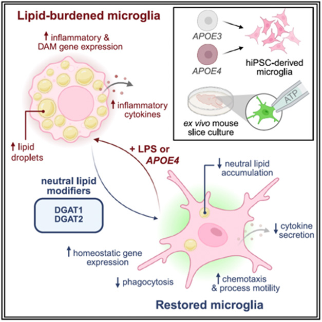
Lipid Central. When provoked by LPS or ApoE4 expression, microglia accumulate lipid droplets (top left) and become activated. Inhibiting triglyceride synthesis with DGAT inhibitors kept the cells calm. [Courtesy of Stephenson et al., Cell Reports, 2025.]
Finally, Stephenson and colleagues noticed that DGAT inhibition triggered an uptick in genes involved in cell motility in ApoE4 microglia. To investigate, the scientists compared how quickly microglia in brain slice cultures re-oriented their processes toward a drop of ATP delivered from a pipette, an established means to measure microglial motility. ApoE4 microglia responded more sluggishly than did ApoE3 microglia. DGAT inhibition corrected this, bringing ApoE4 microglia up to speed.
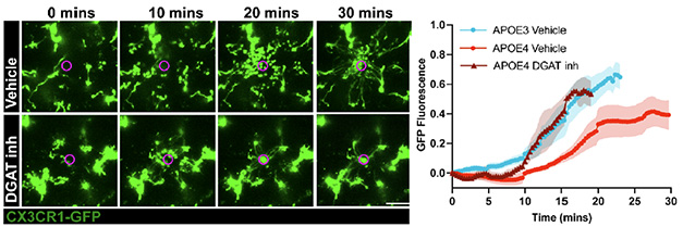
Mobilized. ApoE4 microglia (green) directed their processes toward an ATP stimulus (purple) more rapidly in the presence of DGAT inhibitors (bottom). On right, arrival of microglial processes at ATP stimulus over time. [Courtesy of Stephenson et al., Cell Reports, 2025.]
In all, the findings placed triglyceride metabolism upstream of microglial activation, and suggest that blocking triglyceride production may counteract detrimental effects of ApoE4.
As for how this works, Narayan’s group is investigating angles. For one, microglia may require a large supply of lipids to quickly orient their extensive processes, which after all are lipid bi-layers, toward a given stimulus. Lipids are also the prime source for prostaglandins, inflammatory mediators released by activated microglia. How triglycerides affect the expression of inflammatory genes is less clear, although Narayan does not believe lipid droplets are a source of ATP in microglia, as has recently been reported in neurons (Jul 2025 news).—Jessica Shugart
References
Mutations Citations
News Citations
- Newly Identified Microglia Contain Lipid Droplets, Harm Brain
- Droplets of Unsaturated Fats Burden Human ApoE4 Astrocytes
- Expunging Microglial Lipid Droplets Boosts Phagocytosis
- Paper Alert: APOE4 Packs on Lipid Droplets in Microglia
- Fueled by Fat: Lipid Droplets a Critical Energy Source for Synapses
Paper Citations
- Alzheimer A. Über eine eigenartige Erkrankung der Hirnrinde. Allgemeine Zeitschrift fur Psychiatrie und Psychisch-gerichtliche Medizin. 1907 Jan;64:146-8.
Further Reading
No Available Further Reading
Primary Papers
- Stephenson RA, Sepulveda J, Johnson KR, Lita A, Gopalakrishnan J, Acri DJ, Beilina A, Cheng L, Yang LG, Root JT, Ward ME, Combs C, Skarnes WC, Cookson MR, Shih HY, Larion M, Rebeck GW, Narayan PS. Triglyceride metabolism controls inflammation and microglial phenotypes associated with APOE4. Cell Rep. 2025 Jul 22;44(7):115961. Epub 2025 Jul 10 PubMed.
Annotate
To make an annotation you must Login or Register.
Comments
University of Pennsylvania
University of Pennsylvania
This is a very exciting study. Stephenson et al. rigorously demonstrate that triglyceride (TG) lipid droplets (LDs) play a critical role in the production of damaging cytokines, such as TNF-α and IL1β, in iPSC-derived microglia. The authors do this using multiple iPSC lines and differentiation methods, as well as through both pharmacological and genetic perturbation of DGAT and ACSL1 enzymes. This work further validates many recent studies in mouse models of AD indicating that amyloid induces TG lipid droplets in microglia and that decreasing microglial TG LDs reduces AD pathology in mouse models (Prakash et al., 2025; Wu et al., 2025). Similar to previous reports (Haney et al., 2024), they find that the transcriptional effect of inflammation-induced TG synthesis in iMG is regulated by NF-κB, and importantly, the authors show TG reduction inhibits NF-κB nuclear translocation.
Stephenson et al. further demonstrate these LD phenotypes are related to the APOE4 genotype by showing elevated LDs in isogenic APOE4 induced MG, confirming previous findings that associated APOE4 with TG filled LDs (Victor et al., 2022; Liu et al., 2023; Haney et al. 2024; Friday et al., 2025) and highlighting a cell-autonomous role of the APOE4 genotype in microglia. Importantly, they show that lowering TGs in APOE4 microglia through DGAT inhibition rescues APOE4-related defects in motility in mouse brain slices. These findings give further support to microglial triglyceride lipid droplet accumulation as an important new drug target in AD, a target strongly supported by human genetics but for which there is no therapeutic.
One somewhat paradoxical finding was that inhibition of ATGL, a lipase essential for the catabolism of TGs from LDs, had similar effects on cytokine release and phagocytosis as DGAT inhibition. This would suggest that both LD biosynthesis and catabolism may be necessary for microglial activation in response to LPS. This raises the question of what is the mechanistic link between TG catabolism and microglial response to LPS? This is also very exciting in the context of recent findings that APOE is a lipid droplet-associated protein and that APOE variants modify LD formation and size (Windham et al., 2024). In future studies, it may be interesting to investigate this TG catabolism finding further in the context of the APOE4 genotype, or in animal models, to determine if this is exclusive to LPS stimulation or is a broader aspect of microglial activation in neurodegeneration.
References:
Prakash P, Manchanda P, Paouri E, Bisht K, Sharma K, Rajpoot J, Wendt V, Hossain A, Wijewardhane PR, Randolph CE, Chen Y, Stanko S, Gasmi N, Gjojdeshi A, Card S, Fine J, Jethava KP, Clark MG, Dong B, Ma S, Crockett A, Thayer EA, Nicolas M, Davis R, Hardikar D, Allende D, Prayson RA, Zhang C, Davalos D, Chopra G. Amyloid-β induces lipid droplet-mediated microglial dysfunction via the enzyme DGAT2 in Alzheimer's disease. Immunity. 2025 Jun 10;58(6):1536-1552.e8. Epub 2025 May 19 PubMed.
Wu X, Miller JA, Lee BT, Wang Y, Ruedl C. Reducing microglial lipid load enhances β amyloid phagocytosis in an Alzheimer's disease mouse model. Sci Adv. 2025 Feb 7;11(6):eadq6038. Epub 2025 Feb 5 PubMed.
Haney MS, Pálovics R, Munson CN, Long C, Johansson PK, Yip O, Dong W, Rawat E, West E, Schlachetzki JC, Tsai A, Guldner IH, Lamichhane BS, Smith A, Schaum N, Calcuttawala K, Shin A, Wang YH, Wang C, Koutsodendris N, Serrano GE, Beach TG, Reiman EM, Glass CK, Abu-Remaileh M, Enejder A, Huang Y, Wyss-Coray T. APOE4/4 is linked to damaging lipid droplets in Alzheimer's disease microglia. Nature. 2024 Apr;628(8006):154-161. Epub 2024 Mar 13 PubMed.
Victor MB, Leary N, Luna X, Meharena HS, Scannail AN, Bozzelli PL, Samaan G, Murdock MH, von Maydell D, Effenberger AH, Cerit O, Wen HL, Liu L, Welch G, Bonner M, Tsai LH. Lipid accumulation induced by APOE4 impairs microglial surveillance of neuronal-network activity. Cell Stem Cell. 2022 Aug 4;29(8):1197-1212.e8. PubMed.
Liu CC, Wang N, Chen Y, Inoue Y, Shue F, Ren Y, Wang M, Qiao W, Ikezu TC, Li Z, Zhao J, Martens Y, Doss SV, Rosenberg CL, Jeevaratnam S, Jia L, Raulin AC, Qi F, Zhu Y, Alnobani A, Knight J, Chen Y, Linares C, Kurti A, Fryer JD, Zhang B, Wu LJ, Kim BY, Bu G. Cell-autonomous effects of APOE4 in restricting microglial response in brain homeostasis and Alzheimer's disease. Nat Immunol. 2023 Nov;24(11):1854-1866. Epub 2023 Oct 19 PubMed.
Friday CM, Stephens IO, Smith CT, Lee S, Satish D, Devanney NA, Cohen S, Morganti JM, Gordon SM, Johnson LA. APOE4 reshapes the lipid droplet proteome and modulates microglial inflammatory responses. Neurobiol Dis. 2025 Aug;212:106983. Epub 2025 May 30 PubMed.
Windham IA, Powers AE, Ragusa JV, Wallace ED, Zanellati MC, Williams VH, Wagner CH, White KK, Cohen S. APOE traffics to astrocyte lipid droplets and modulates triglyceride saturation and droplet size. J Cell Biol. 2024 Apr 1;223(4) Epub 2024 Feb 9 PubMed.
Make a Comment
To make a comment you must login or register.