Synapses Found to Vanish on Same Time Scale as Memories
Quick Links
Researchers believe that the brain’s cerebral cortex encodes long-term memories by forming and stabilizing synapse-bearing dendritic spines. What, then, happens in the hippocampus, which stores memories only transiently? The question has gone unanswered partly because the high spine density in the hippocampus makes it difficult to follow the fate of individual synapses. On June 22 in Nature, researchers led by Mark Schnitzer at Stanford University, California, describe a new computational approach that allowed them to calculate the number and dynamics of hippocampal spines during weeks of live imaging in transgenic mice. The data revealed that dendritic spines, unlike their cortical cousins, do not last. The authors estimated the entire population turns over in six weeks or less, roughly matching the timespan of hippocampal-dependent memory in mice. The results support current models of memory storage but also highlight striking differences between the cortex and hippocampus. The techniques described in this paper could be used to address additional questions about hippocampal memory mechanisms, Schnitzer suggested.
Commentators agreed. “This is a technical tour de force,” said Scott Small at Columbia University, New York. Howard Eichenbaum at Boston University called the data exciting. “The observations reveal new information about how memories last or decay, and so open avenues to explore the mechanisms of memory loss in neurological disorders such as Alzheimer’s,” he wrote to Alzforum.
Previous studies indicated that dendrites and spines in the neocortex wax and wane over time, influenced by an animal’s experience (see Dec 2002 news; Jan 2006 news). In live imaging experiments, the neocortex appears to contain a mix of transient and long-lasting dendritic spines, in keeping with its abilities to learn and change as well as store permanent memories (see Holtmaat et al., 2005; Dec 2009 news). In contrast to the relatively sparser spines in cortex, however, many more spines crowd hippocampal tissue. The two-photon microscopy used for live imaging cannot reliably distinguish many of these closely spaced spines, making visual counts unreliable.

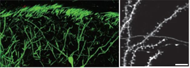
Secret Life of Synapses. Fluorescent pyramidal neurons in the hippocampi of transgenic mice (left panel) allow researchers to visualize changes in dendritic spines (knobby protrusions in right panel). [Courtesy of Attardo et al., Nature.]
To solve the problem, Schnitzer and joint first authors Alessio Attardo and James Fitzgerald first compared two-photon microscopy with the more sensitive stimulated emission depletion (STED) microscopy in hippocampal tissue slices from transgenic Thy1-GFP mice. These animals express GFP in a subset of CA1 pyramidal neurons, allowing these neurons and their spines to be visualized (see image above). About 30 percent of spines that appeared single in two-photon images in fact were shown to be two, or sometimes three, spines close together in STED images. Two-photon data thus underestimates spine density and overestimates spine stability, since in many cases two spines would need to retract for an apparent spine to disappear from the image. The authors put this data into computer simulations of various hypothetical spine densities and kinetics, which they used to develop a mathematical model for correcting the two-photon data.
The authors then implanted microendoscopes into the brains of four Thy1-GFP mice just above the CA1 region of hippocampus. These needle-thin devices contain lenses that relay a signal back to a two-photon microscope. The authors acquired images every three days for three weeks to track spine dynamics, then analyzed the data using their mathematical model. The best fit for the observed data was a model in which 100 percent of spines turned over, with an average lifespan of about 10 days. This would lead to complete replacement of hippocampal spines every three to six weeks, the authors calculated.
Environmental enrichment has been found to increase hippocampal spine density in some studies (see Moser et al., 1994; Rampon et al., 2000). The authors tracked three new mice, first for three weeks in a regular cage and then for 24 days in a cage filled with playthings. However, neither spine density nor turnover changed in the richer environment, belying earlier studies.
Since NMDA glutamate receptors are known to affect synaptic plasticity in both the cortex and hippocampus, the authors investigated the effects of injecting mice with an NMDA receptor blocker twice daily for 18 days. More spines disappeared after treatment, leading to about 12 percent less spine density. The finding suggests that CA1 dendritic spines require working NMDA receptors to survive.
Intriguingly, cortical spines appear to need just the opposite. There, blockade of NMDA receptors has been reported to stabilize spines (e.g., Zuo et al., 2005). Moreover, in adult neocortices more than half of spines seem to be permanent, and those that are transitory have an average lifespan of about five days. To compare cortical and hippocampal spine dynamics on an even footing, Schnitzer and colleagues re-analyzed data from previous cortical imaging studies using their mathematical model. The results confirmed that about 60 percent of cortical spines remained stable over long periods of time. “The data highlight that the two systems have very different manners of memory processing,” Schnitzer told Alzforum. In future studies, he would like to investigate the mechanisms that control spine turnover in the hippocampus, and also relate spine dynamics to memory formation and behavior in mice.
Small noted that the differences in structural plasticity seen in the hippocampus and neocortex track well with the known memory durations in each region. “That’s very elegant. There has always been a question about what cellular mechanism underlies the temporal distinction in memory codes,” he said. Moreover, structural changes occur slowly over days or weeks, and therefore may better reflect the brain changes seen during repeated functional MRI or FDG PET than neuronal activity does, Small suggested. It would be interesting to look for a functional imaging correlate for spine turnover, he added. Such a correlate could allow researchers to study these changes in people.
The technique described here could also be applied to animal models of Alzheimer’s and other memory disorders, researchers agreed. Synapses are lost early in AD (see Jul 2007 conference news; Jun 2012 news). “There might be a number of different mouse models of disease that would have altered spine kinetics,” Schnitzer speculated.—Madolyn Bowman Rogers
References
News Citations
- Dendritic Spine Stability—Not So Black and White—or Is That Green and Yellow?
- Neuronal Plasticity the Arbor Way—Dendritic Remodeling in the Neocortex
- Persistence of Dendrites Leads to Lifelong Memories
- Paris: Synaptic Plasticity and the Mechanism of Alzheimer Disease
- Synaptic Plasticity Falters Early in AD Mice
Paper Citations
- Holtmaat AJ, Trachtenberg JT, Wilbrecht L, Shepherd GM, Zhang X, Knott GW, Svoboda K. Transient and persistent dendritic spines in the neocortex in vivo. Neuron. 2005 Jan 20;45(2):279-91. PubMed.
- Moser MB, Trommald M, Andersen P. An increase in dendritic spine density on hippocampal CA1 pyramidal cells following spatial learning in adult rats suggests the formation of new synapses. Proc Natl Acad Sci U S A. 1994 Dec 20;91(26):12673-5. PubMed.
- Rampon C, Tang YP, Goodhouse J, Shimizu E, Kyin M, Tsien JZ. Enrichment induces structural changes and recovery from nonspatial memory deficits in CA1 NMDAR1-knockout mice. Nat Neurosci. 2000 Mar;3(3):238-44. PubMed.
- Zuo Y, Lin A, Chang P, Gan WB. Development of long-term dendritic spine stability in diverse regions of cerebral cortex. Neuron. 2005 Apr 21;46(2):181-9. PubMed.
Further Reading
News
- Prefrontal Hook-Up Required for New Memories in the Hippocampus
- Could Silencing Neuroligin-1 Drive Synaptic Loss in AD?
- The Guardians of Forever: Forming and Keeping Memories
- Mnemonic Models—Flies, Mice, and Rats Reveal Pathways to Memory
- Myosin and Memory: Motor Protein’s Major Role in Synapse Remodeling
- Do Synaptic Tags Code Memories for Storage?
Primary Papers
- Attardo A, Fitzgerald JE, Schnitzer MJ. Impermanence of dendritic spines in live adult CA1 hippocampus. Nature. 2015 Jun 22; PubMed.
Annotate
To make an annotation you must Login or Register.

Comments
No Available Comments
Make a Comment
To make a comment you must login or register.